 Movie
Movie Controller


+ Open data

- Basic information
| Entry | Database: PDB / ID: 1jct | ||||||
|---|---|---|---|---|---|---|---|
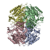
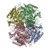
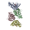
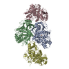
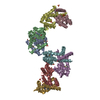
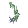
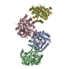
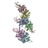
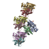
| Title | Glucarate Dehydratase, N341L mutant Orthorhombic Form | ||||||
 Components Components | Glucarate Dehydratase | ||||||
 Keywords Keywords | LYASE / alpha/beta barrel / Enolase superfamily | ||||||
| Function / homology |  Function and homology information Function and homology informationD-glucarate catabolic process / glucarate dehydratase / glucarate dehydratase activity / magnesium ion binding Similarity search - Function | ||||||
| Biological species |  | ||||||
| Method |  X-RAY DIFFRACTION / SYNCHROTRON / MOLECULAR REPLACEMENT / Resolution: 2.75 Å X-RAY DIFFRACTION / SYNCHROTRON / MOLECULAR REPLACEMENT / Resolution: 2.75 Å | ||||||
 Authors Authors | Gulick, A.M. / Hubbard, B.K. / Gerlt, J.A. / Rayment, I. | ||||||
 Citation Citation | Journal: Biochemistry / Year: 2001 Title: Evolution of enzymatic activities in the enolase superfamily: identification of the general acid catalyst in the active site of D-glucarate dehydratase from Escherichia coli. Authors: Gulick, A.M. / Hubbard, B.K. / Gerlt, J.A. / Rayment, I. #1: Journal: Biochemistry / Year: 2000Title: Evolution of Enzymatic Active Sites in the Enolase Superfamily: crystallographic and mutagenesis studies of the reaction catalyzed by D-glucarate dehydratase from Escherichia coli. Authors: Gulick, A.M. / Hubbard, B.K. / Gerlt, J.A. / Rayment, I. | ||||||
| History |
|
- Structure visualization
| Structure viewer | Molecule: MolmilJmol/JSmol |
|---|
- Downloads & links
-Download
| PDBx/mmCIF format | 1jct.cif.gz | 179.9 KB | Display | PDBx/mmCIF format |
|---|---|---|---|---|
| PDB format | pdb1jct.ent.gz | 142.8 KB | Display | PDB format |
| PDBx/mmJSON format | 1jct.json.gz | Tree view | PDBx/mmJSON format | |
| Others |  Other downloads Other downloads |
-Validation report
| Arichive directory | https://data.pdbj.org/pub/pdb/validation_reports/jc/1jctftp://data.pdbj.org/pub/pdb/validation_reports/jc/1jct | HTTPS FTP |
|---|
-Related structure data
| Related structure data |  1jdfC  1ec8S S: Starting model for refinement C: citing same article ( |
|---|---|
| Similar structure data |


-Links
 PDBj
PDBj

- Assembly
| Deposited unit | 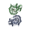
| ||||||||
|---|---|---|---|---|---|---|---|---|---|
| 1 | 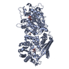
| ||||||||
| 2 | 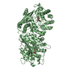
| ||||||||
| Unit cell |
| ||||||||
| Components on special symmetry positions |
|
-Components
| #1: Protein | Mass: 49195.910 Da / Num. of mol.: 2 / Mutation: N341L Source method: isolated from a genetically manipulated source Source: (gene. exp.) References: UniProt: P76637, UniProt: P0AES2*PLUS, glucarate dehydratase #2: Chemical |   Mass: 24.305 Da / Num. of mol.: 2 / Source method: obtained synthetically / Formula: Mg Mass: 24.305 Da / Num. of mol.: 2 / Source method: obtained synthetically / Formula: Mg#3: Chemical |   Mass: 208.123 Da / Num. of mol.: 2 / Source method: obtained synthetically / Formula: C6H8O8 Mass: 208.123 Da / Num. of mol.: 2 / Source method: obtained synthetically / Formula: C6H8O8#4: Chemical |   Mass: 60.095 Da / Num. of mol.: 2 / Source method: obtained synthetically / Formula: C3H8O Mass: 60.095 Da / Num. of mol.: 2 / Source method: obtained synthetically / Formula: C3H8O#5: Water | ChemComp-HOH / |  Mass: 18.015 Da / Num. of mol.: 96 / Source method: isolated from a natural source / Formula: H2O Mass: 18.015 Da / Num. of mol.: 96 / Source method: isolated from a natural source / Formula: H2O |
|---|
-Experimental details
-Experiment
| Experiment | Method: X-RAY DIFFRACTION / Number of used crystals: 1 |
|---|
- Sample preparation
| Crystal | Density Matthews: 2.6 Å3/Da / Density % sol: 52.64 % | ||||||||||||||||||||||||||||||||||||||||||||||||||||||||
|---|---|---|---|---|---|---|---|---|---|---|---|---|---|---|---|---|---|---|---|---|---|---|---|---|---|---|---|---|---|---|---|---|---|---|---|---|---|---|---|---|---|---|---|---|---|---|---|---|---|---|---|---|---|---|---|---|---|
| Crystal grow | Temperature: 277 K / Method: vapor diffusion, hanging drop / pH: 8 Details: Peg5000 monomethylether, 50 mM MgCl, 5% isopropanol, 50 mM HEPPS, pH 8.0, VAPOR DIFFUSION, HANGING DROP, temperature 277K | ||||||||||||||||||||||||||||||||||||||||||||||||||||||||
| Crystal grow | *PLUS Temperature: 4 ℃ / Method: batch method / Details: used macroseeding | ||||||||||||||||||||||||||||||||||||||||||||||||||||||||
| Components of the solutions | *PLUS
|
-Data collection
| Diffraction | Mean temperature: 113 K |
|---|---|
| Diffraction source | Source: SYNCHROTRON / Site: APS  / Beamline: 19-BM / Beamline: 19-BM |
| Detector | Type: CUSTOM-MADE / Detector: CCD / Date: Jun 23, 2000 |
| Radiation | Monochromator: 0.1 mm / Protocol: SINGLE WAVELENGTH / Monochromatic (M) / Laue (L): M / Scattering type: x-ray |
| Radiation wavelength | Relative weight: 1 |
| Reflection | Resolution: 2.75→30 Å / Num. all: 27057 / Num. obs: 24649 / % possible obs: 91.1 % / Observed criterion σ(F): -3 / Observed criterion σ(I): -3 / Redundancy: 4.1 % / Biso Wilson estimate: 68.7 Å2 / Rmerge(I) obs: 0.071 / Net I/σ(I): 10.5 |
| Reflection shell | Resolution: 2.75→2.85 Å / Redundancy: 4 % / Rmerge(I) obs: 0.394 / % possible all: 93.5 |
| Reflection | *PLUS Lowest resolution: 30 Å / Num. measured all: 100153 |
| Reflection shell | *PLUS % possible obs: 93.5 % |
- Processing
| Software |
| |||||||||||||||||||||
|---|---|---|---|---|---|---|---|---|---|---|---|---|---|---|---|---|---|---|---|---|---|---|
| Refinement | Method to determine structure: MOLECULAR REPLACEMENT Starting model: 1EC8 Resolution: 2.75→30 Å / σ(F): 0 / σ(I): 0 / Stereochemistry target values: Engh & Huber
| |||||||||||||||||||||
| Displacement parameters | Biso mean: 55 Å2
| |||||||||||||||||||||
| Refine analyze |
| |||||||||||||||||||||
| Refinement step | Cycle: LAST / Resolution: 2.75→30 Å
| |||||||||||||||||||||
| Refine LS restraints |
| |||||||||||||||||||||
| LS refinement shell | Resolution: 2.75→2.92 Å / Rfactor Rfree error: 0.03
| |||||||||||||||||||||
| Software | *PLUS Name: CNS / Classification: refinement | |||||||||||||||||||||
| Refinement | *PLUS Lowest resolution: 30 Å / σ(F): 0 / Rfactor obs: 0.213 | |||||||||||||||||||||
| Solvent computation | *PLUS | |||||||||||||||||||||
| Displacement parameters | *PLUS Biso mean: 55 Å2 | |||||||||||||||||||||
| Refine LS restraints | *PLUS
| |||||||||||||||||||||
| LS refinement shell | *PLUS Rfactor Rfree: 0.449 / Rfactor Rwork: 0.347 |
