 Movie
Movie Controller

[English] 日本語
 Yorodumi
- PDB-1j7l: Crystal Structure of 3',5"-Aminoglycoside Phosphotransferase Type... -
+ Open data

- Basic information
| Entry | Database: PDB / ID: 1j7l | ||||||
|---|---|---|---|---|---|---|---|
| Title | Crystal Structure of 3',5"-Aminoglycoside Phosphotransferase Type IIIa ADP Complex | ||||||
 Components Components | AMINOGLYCOSIDE 3'-PHOSPHOTRANSFERASE | ||||||
 Keywords Keywords | TRANSFERASE / antibiotic resistance / kinase / ATP-binding | ||||||
| Function / homology |  Function and homology information Function and homology informationkanamycin kinase / kanamycin kinase activity / response to antibiotic / ATP binding Similarity search - Function | ||||||
| Biological species |   Enterococcus faecalis (bacteria) Enterococcus faecalis (bacteria) | ||||||
| Method |  X-RAY DIFFRACTION / SYNCHROTRON / MAD / Resolution: 2.2 Å X-RAY DIFFRACTION / SYNCHROTRON / MAD / Resolution: 2.2 Å | ||||||
 Authors Authors | Burk, D.L. / Hon, W.C. / Leung, A.K.-W. / Berghuis, A.M. | ||||||
 Citation Citation | Journal: Biochemistry / Year: 2001 Title: Structural analyses of nucleotide binding to an aminoglycoside phosphotransferase. Authors: Burk, D.L. / Hon, W.C. / Leung, A.K. / Berghuis, A.M. #1: Journal: Cell(Cambridge,Mass.) / Year: 1997Title: Structure of an Enzyme Required for Antibiotic Resistance Reveals Homology to Eukaryotic Protein Kinases Authors: Hon, W.C. / McKay, G.A. / Thompson, P.R. / Sweet, R.M. / Yang, D.S.C. / Wright, G.D. / Berghuis, A.M. | ||||||
| History |
|
- Structure visualization
| Structure viewer | Molecule: MolmilJmol/JSmol |
|---|
- Downloads & links
-Download
| PDBx/mmCIF format | 1j7l.cif.gz | 129.5 KB | Display | PDBx/mmCIF format |
|---|---|---|---|---|
| PDB format | pdb1j7l.ent.gz | 100.6 KB | Display | PDB format |
| PDBx/mmJSON format | 1j7l.json.gz | Tree view | PDBx/mmJSON format | |
| Others |  Other downloads Other downloads |
-Validation report
| Arichive directory | https://data.pdbj.org/pub/pdb/validation_reports/j7/1j7lftp://data.pdbj.org/pub/pdb/validation_reports/j7/1j7l | HTTPS FTP |
|---|
-Related structure data


-Links
 PDBj
PDBj
- Assembly
| Deposited unit | 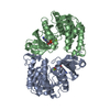
| ||||||||
|---|---|---|---|---|---|---|---|---|---|
| 1 |
| ||||||||
| Unit cell |
|
-Components
| #1: Protein | Mass: 31012.045 Da / Num. of mol.: 2 Source method: isolated from a genetically manipulated source Source: (gene. exp.) Enterococcus faecalis (bacteria) / Plasmid: pPCRG6 / Species (production host): Escherichia coli / Production host: #2: Chemical | ChemComp-MG /   Mass: 24.305 Da / Num. of mol.: 4 / Source method: obtained synthetically / Formula: Mg Mass: 24.305 Da / Num. of mol.: 4 / Source method: obtained synthetically / Formula: Mg#3: Chemical |   Mass: 427.201 Da / Num. of mol.: 2 / Source method: obtained synthetically / Formula: C10H15N5O10P2 / Comment: ADP, energy-carrying molecule*YM Mass: 427.201 Da / Num. of mol.: 2 / Source method: obtained synthetically / Formula: C10H15N5O10P2 / Comment: ADP, energy-carrying molecule*YM#4: Water | ChemComp-HOH / |  Mass: 18.015 Da / Num. of mol.: 297 / Source method: isolated from a natural source / Formula: H2O Mass: 18.015 Da / Num. of mol.: 297 / Source method: isolated from a natural source / Formula: H2OHas protein modification | Y | |
|---|
-Experimental details
-Experiment
| Experiment | Method: X-RAY DIFFRACTION / Number of used crystals: 1 |
|---|
- Sample preparation
| Crystal | Density Matthews: 2.4 Å3/Da / Density % sol: 48.74 % | |||||||||||||||||||||||||
|---|---|---|---|---|---|---|---|---|---|---|---|---|---|---|---|---|---|---|---|---|---|---|---|---|---|---|
| Crystal grow | Temperature: 298 K / Method: vapor diffusion, hanging drop / pH: 6.5 Details: PEG 8000, magnesium acetate, sodium cacodylate, adenosine-5'-triphosphate , pH 6.5, VAPOR DIFFUSION, HANGING DROP, temperature 298K | |||||||||||||||||||||||||
| Crystal grow | *PLUS Details: Hon, W.C., (1997) Cell (Cambridge,Mass.), 89, 887. | |||||||||||||||||||||||||
| Components of the solutions | *PLUS
|
-Data collection
| Diffraction | Mean temperature: 110 K |
|---|---|
| Diffraction source | Source: SYNCHROTRON / Site: NSLS  / Beamline: X12C / Wavelength: 1.05 Å / Beamline: X12C / Wavelength: 1.05 Å |
| Detector | Type: MARRESEARCH / Detector: IMAGE PLATE |
| Radiation | Protocol: SINGLE WAVELENGTH / Monochromatic (M) / Laue (L): M / Scattering type: x-ray |
| Radiation wavelength | Wavelength: 1.05 Å / Relative weight: 1 |
| Reflection | Resolution: 2.2→37.47 Å / Num. all: 95428 / Num. obs: 95428 / % possible obs: 96.4 % / Observed criterion σ(F): 0 / Observed criterion σ(I): 0 / Redundancy: 3.1 % / Biso Wilson estimate: 17.6 Å2 / Rmerge(I) obs: 0.055 / Net I/σ(I): 13.9 |
| Reflection shell | Resolution: 2.2→2.34 Å / Rmerge(I) obs: 0.213 / Mean I/σ(I) obs: 4.7 / Num. unique all: 4389 / % possible all: 95.9 |
| Reflection | *PLUS Num. obs: 30902 / Num. measured all: 95428 |
| Reflection shell | *PLUS Lowest resolution: 2.28 Å / % possible obs: 95.9 % |
- Processing
| Software |
| ||||||||||||||||||||||||||||||||||||||||||||
|---|---|---|---|---|---|---|---|---|---|---|---|---|---|---|---|---|---|---|---|---|---|---|---|---|---|---|---|---|---|---|---|---|---|---|---|---|---|---|---|---|---|---|---|---|---|
| Refinement | Method to determine structure: MAD / Resolution: 2.2→37.47 Å / Rfactor Rfree error: 0.005 / Data cutoff high absF: 44702935.64 / Data cutoff low absF: 0 / Isotropic thermal model: RESTRAINED / Cross valid method: THROUGHOUT / σ(F): 0 / σ(I): 0 / Stereochemistry target values: Engh & Huber
| ||||||||||||||||||||||||||||||||||||||||||||
| Solvent computation | Solvent model: FLAT MODEL / Bsol: 24.45 Å2 / ksol: 0.3807 e/Å3 | ||||||||||||||||||||||||||||||||||||||||||||
| Displacement parameters | Biso mean: 22.9 Å2
| ||||||||||||||||||||||||||||||||||||||||||||
| Refine analyze |
| ||||||||||||||||||||||||||||||||||||||||||||
| Refinement step | Cycle: LAST / Resolution: 2.2→37.47 Å
| ||||||||||||||||||||||||||||||||||||||||||||
| Refine LS restraints |
| ||||||||||||||||||||||||||||||||||||||||||||
| LS refinement shell | Resolution: 2.2→2.34 Å / Rfactor Rfree error: 0.014 / Total num. of bins used: 6
| ||||||||||||||||||||||||||||||||||||||||||||
| Software | *PLUS Name: CNS / Version: 1 / Classification: refinement | ||||||||||||||||||||||||||||||||||||||||||||
| Refinement | *PLUS σ(F): 0 / % reflection Rfree: 10 % / Rfactor obs: 0.22 / Rfactor Rwork: 0.22 | ||||||||||||||||||||||||||||||||||||||||||||
| Solvent computation | *PLUS | ||||||||||||||||||||||||||||||||||||||||||||
| Displacement parameters | *PLUS Biso mean: 22.9 Å2 | ||||||||||||||||||||||||||||||||||||||||||||
| Refine LS restraints | *PLUS
| ||||||||||||||||||||||||||||||||||||||||||||
| LS refinement shell | *PLUS Rfactor Rfree: 0.311 / % reflection Rfree: 10.3 % / Rfactor Rwork: 0.241 |
