 Movie
Movie Controller


+ Open data

- Basic information
| Entry | Database: PDB / ID: 1j74 | ||||||
|---|---|---|---|---|---|---|---|
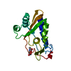
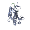
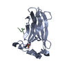
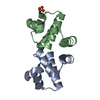
| Title | Crystal Structure of Mms2 | ||||||
 Components Components | MMS2 | ||||||
 Keywords Keywords | UNKNOWN FUNCTION / Mms2 / Uev / Ubiquitin / Ubc / DNA repair | ||||||
| Function / homology |  Function and homology information Function and homology informationerror-free postreplication DNA repair / UBC13-MMS2 complex / ubiquitin conjugating enzyme complex / positive regulation of protein K63-linked ubiquitination / DNA double-strand break processing / DNA damage tolerance / positive regulation of double-strand break repair / protein K63-linked ubiquitination / regulation of DNA repair / Nonhomologous End-Joining (NHEJ) ...error-free postreplication DNA repair / UBC13-MMS2 complex / ubiquitin conjugating enzyme complex / positive regulation of protein K63-linked ubiquitination / DNA double-strand break processing / DNA damage tolerance / positive regulation of double-strand break repair / protein K63-linked ubiquitination / regulation of DNA repair / Nonhomologous End-Joining (NHEJ) / G2/M DNA damage checkpoint / Formation of Incision Complex in GG-NER / protein polyubiquitination / Antigen processing: Ubiquitination & Proteasome degradation / E3 ubiquitin ligases ubiquitinate target proteins / Recruitment and ATM-mediated phosphorylation of repair and signaling proteins at DNA double strand breaks / Processing of DNA double-strand break ends / protein ubiquitination / extracellular exosome / nucleoplasm / nucleus / cytoplasm Similarity search - Function | ||||||
| Biological species |  Homo sapiens (human) Homo sapiens (human) | ||||||
| Method |  X-RAY DIFFRACTION / MOLECULAR REPLACEMENT / Resolution: 1.9 Å X-RAY DIFFRACTION / MOLECULAR REPLACEMENT / Resolution: 1.9 Å | ||||||
 Authors Authors | Moraes, T.F. / Edwards, R.A. / McKenna, S. / Pastushok, L. / Xiao, W. / Glover, J.N.M. / Ellison, M.J. | ||||||
 Citation Citation | Journal: Nat.Struct.Biol. / Year: 2001 Title: Crystal structure of the human ubiquitin conjugating enzyme complex, hMms2-hUbc13. Authors: Moraes, T.F. / Edwards, R.A. / McKenna, S. / Pastushok, L. / Xiao, W. / Glover, J.N. / Ellison, M.J. | ||||||
| History |
|
- Structure visualization
| Structure viewer | Molecule: MolmilJmol/JSmol |
|---|
- Downloads & links
-Download
| PDBx/mmCIF format | 1j74.cif.gz | 41 KB | Display | PDBx/mmCIF format |
|---|---|---|---|---|
| PDB format | pdb1j74.ent.gz | 28.5 KB | Display | PDB format |
| PDBx/mmJSON format | 1j74.json.gz | Tree view | PDBx/mmJSON format | |
| Others |  Other downloads Other downloads |
-Validation report
| Arichive directory | https://data.pdbj.org/pub/pdb/validation_reports/j7/1j74ftp://data.pdbj.org/pub/pdb/validation_reports/j7/1j74 | HTTPS FTP |
|---|
-Related structure data
| Related structure data |  1j7dSC S: Starting model for refinement C: citing same article ( |
|---|---|
| Similar structure data |


-Links
 PDBj
PDBj


- Assembly
| Deposited unit | 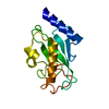
| ||||||||
|---|---|---|---|---|---|---|---|---|---|
| 1 |
| ||||||||
| Unit cell |
|
-Components
| #1: Protein | Mass: 16380.731 Da / Num. of mol.: 1 Source method: isolated from a genetically manipulated source Source: (gene. exp.) Homo sapiens (human) / Production host:  |
|---|---|
| #2: Water | ChemComp-HOH /  Mass: 18.015 Da / Num. of mol.: 99 / Source method: isolated from a natural source / Formula: H2O Mass: 18.015 Da / Num. of mol.: 99 / Source method: isolated from a natural source / Formula: H2O |
-Experimental details
-Experiment
| Experiment | Method: X-RAY DIFFRACTION / Number of used crystals: 1 |
|---|
- Sample preparation
| Crystal | Density Matthews: 1.9 Å3/Da / Density % sol: 35.22 % | ||||||||||||||||||||||||||||||||||||||||||||||||||||||
|---|---|---|---|---|---|---|---|---|---|---|---|---|---|---|---|---|---|---|---|---|---|---|---|---|---|---|---|---|---|---|---|---|---|---|---|---|---|---|---|---|---|---|---|---|---|---|---|---|---|---|---|---|---|---|---|
| Crystal grow | Temperature: 298 K / Method: vapor diffusion, hanging drop / pH: 6.5 Details: 16% PEG 6000, 25mM NaCl, 100mM citrate, pH 6.5, VAPOR DIFFUSION, HANGING DROP, temperature 298K | ||||||||||||||||||||||||||||||||||||||||||||||||||||||
| Crystal grow | *PLUS Temperature: 20-24 ℃ / pH: 7.5 | ||||||||||||||||||||||||||||||||||||||||||||||||||||||
| Components of the solutions | *PLUS
|
-Data collection
| Diffraction | Mean temperature: 105 K |
|---|---|
| Diffraction source | Source: ROTATING ANODE / Type: RIGAKU RUH3R / Wavelength: 1.5418 Å |
| Detector | Type: RIGAKU RAXIS IV / Detector: IMAGE PLATE / Date: Jul 21, 2000 / Details: mirrors |
| Radiation | Protocol: SINGLE WAVELENGTH / Monochromatic (M) / Laue (L): M / Scattering type: x-ray |
| Radiation wavelength | Wavelength: 1.5418 Å / Relative weight: 1 |
| Reflection | Resolution: 1.9→20 Å / Num. all: 9995 / Num. obs: 9973 / % possible obs: 97.9 % / Redundancy: 5.35 % / Biso Wilson estimate: 19.7 Å2 / Rmerge(I) obs: 0.045 / Rsym value: 4.5 / Net I/σ(I): 37.1 |
| Reflection shell | Resolution: 1.9→1.93 Å / Redundancy: 3 % / Rmerge(I) obs: 0.129 / Mean I/σ(I) obs: 20 / Num. unique all: 445 / Rsym value: 12.9 / % possible all: 87.6 |
| Reflection | *PLUS Lowest resolution: 20 Å / Num. measured all: 53554 |
- Processing
| Software |
| |||||||||||||||||||||||||
|---|---|---|---|---|---|---|---|---|---|---|---|---|---|---|---|---|---|---|---|---|---|---|---|---|---|---|
| Refinement | Method to determine structure: MOLECULAR REPLACEMENT Starting model: PDB entry 1J7D Resolution: 1.9→20 Å / Stereochemistry target values: CNS template
| |||||||||||||||||||||||||
| Refinement step | Cycle: LAST / Resolution: 1.9→20 Å
| |||||||||||||||||||||||||
| Refine LS restraints |
| |||||||||||||||||||||||||
| LS refinement shell | Resolution: 1.9→1.98 Å
| |||||||||||||||||||||||||
| Software | *PLUS Name: CNS / Classification: refinement | |||||||||||||||||||||||||
| Refinement | *PLUS Highest resolution: 1.9 Å / Lowest resolution: 20 Å / % reflection Rfree: 5 % / Rfactor obs: 0.213 | |||||||||||||||||||||||||
| Solvent computation | *PLUS | |||||||||||||||||||||||||
| Displacement parameters | *PLUS |
