 Movie
Movie Controller

[English] 日本語
 Yorodumi
- PDB-1j54: Structure of the N-terminal exonuclease domain of the epsilon sub... -
+ Open data

- Basic information
| Entry | Database: PDB / ID: 1j54 | ||||||
|---|---|---|---|---|---|---|---|
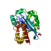
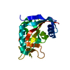
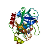
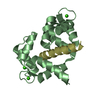
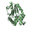
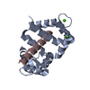
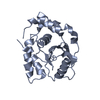
| Title | Structure of the N-terminal exonuclease domain of the epsilon subunit of E.coli DNA polymerase III at pH 5.8 | ||||||
 Components Components | DNA polymerase III, epsilon chain | ||||||
 Keywords Keywords | TRANSFERASE / DNA polymerase proofreading domain | ||||||
| Function / homology |  Function and homology information Function and homology informationDNA polymerase III, core complex / DNA replication proofreading / DNA polymerase III complex / lagging strand elongation / replisome / exonuclease activity / leading strand elongation / 3'-5' exonuclease activity / DNA-templated DNA replication / DNA-directed DNA polymerase ...DNA polymerase III, core complex / DNA replication proofreading / DNA polymerase III complex / lagging strand elongation / replisome / exonuclease activity / leading strand elongation / 3'-5' exonuclease activity / DNA-templated DNA replication / DNA-directed DNA polymerase / DNA-directed DNA polymerase activity / DNA binding / metal ion binding / cytosol Similarity search - Function | ||||||
| Biological species |  | ||||||
| Method |  X-RAY DIFFRACTION / SYNCHROTRON / Resolution: 1.7 Å X-RAY DIFFRACTION / SYNCHROTRON / Resolution: 1.7 Å | ||||||
 Authors Authors | Hamdan, S. / Carr, P.D. / Brown, S.E. / Ollis, D.L. / Dixon, N.E. | ||||||
 Citation Citation | Journal: Structure / Year: 2002 Title: Structural Basis for Proofreading during Replication of the Escherichia coli Chromosome Authors: Hamdan, S. / Carr, P.D. / Brown, S.E. / Ollis, D.L. / Dixon, N.E. | ||||||
| History |
|
- Structure visualization
| Structure viewer | Molecule: MolmilJmol/JSmol |
|---|
- Downloads & links
-Download
| PDBx/mmCIF format | 1j54.cif.gz | 54.9 KB | Display | PDBx/mmCIF format |
|---|---|---|---|---|
| PDB format | pdb1j54.ent.gz | 38.8 KB | Display | PDB format |
| PDBx/mmJSON format | 1j54.json.gz | Tree view | PDBx/mmJSON format | |
| Others |  Other downloads Other downloads |
-Validation report
| Arichive directory | https://data.pdbj.org/pub/pdb/validation_reports/j5/1j54ftp://data.pdbj.org/pub/pdb/validation_reports/j5/1j54 | HTTPS FTP |
|---|
-Related structure data


-Links
 PDBj
PDBj- Assembly
| Deposited unit | 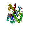
| |||||||||||||||
|---|---|---|---|---|---|---|---|---|---|---|---|---|---|---|---|---|
| 1 |
| |||||||||||||||
| Unit cell |
| |||||||||||||||
| Components on special symmetry positions |
|
-Components
| #1: Protein | Mass: 20741.689 Da / Num. of mol.: 1 / Fragment: N-terminal exonuclease domain (residues 1-186) Source method: isolated from a genetically manipulated source Source: (gene. exp.)   Enterobacteria phage T7 (virus) / References: UniProt: P03007, DNA-directed DNA polymerase Enterobacteria phage T7 (virus) / References: UniProt: P03007, DNA-directed DNA polymerase | ||||||
|---|---|---|---|---|---|---|---|
| #2: Chemical |   Mass: 54.938 Da / Num. of mol.: 2 / Source method: obtained synthetically / Formula: Mn Mass: 54.938 Da / Num. of mol.: 2 / Source method: obtained synthetically / Formula: Mn#3: Chemical | ChemComp-TMP / |   Mass: 322.208 Da / Num. of mol.: 1 / Source method: obtained synthetically / Formula: C10H15N2O8P Mass: 322.208 Da / Num. of mol.: 1 / Source method: obtained synthetically / Formula: C10H15N2O8P#4: Chemical |   Mass: 62.068 Da / Num. of mol.: 3 / Source method: obtained synthetically / Formula: C2H6O2 Mass: 62.068 Da / Num. of mol.: 3 / Source method: obtained synthetically / Formula: C2H6O2#5: Water | ChemComp-HOH / |  Mass: 18.015 Da / Num. of mol.: 225 / Source method: isolated from a natural source / Formula: H2O Mass: 18.015 Da / Num. of mol.: 225 / Source method: isolated from a natural source / Formula: H2O |
-Experimental details
-Experiment
| Experiment | Method: X-RAY DIFFRACTION / Number of used crystals: 1 |
|---|
- Sample preparation
| Crystal | Density Matthews: 2.47 Å3/Da / Density % sol: 50.29 % | ||||||||||||||||||||||||||||||||||||||||||||||||||||||||
|---|---|---|---|---|---|---|---|---|---|---|---|---|---|---|---|---|---|---|---|---|---|---|---|---|---|---|---|---|---|---|---|---|---|---|---|---|---|---|---|---|---|---|---|---|---|---|---|---|---|---|---|---|---|---|---|---|---|
| Crystal grow | Temperature: 277 K / Method: vapor diffusion, hanging drop / pH: 5.8 Details: PEG 8000, magnesium sulfate, cacodylate, pH 5.8, VAPOR DIFFUSION, HANGING DROP, temperature 277K | ||||||||||||||||||||||||||||||||||||||||||||||||||||||||
| Crystal grow | *PLUS Temperature: 4 ℃ / pH: 7.5 / Details: Hamdan, S., (2000) J.Struct.Biol., 131, 164. | ||||||||||||||||||||||||||||||||||||||||||||||||||||||||
| Components of the solutions | *PLUS
|
-Data collection
| Diffraction | Mean temperature: 100 K |
|---|---|
| Diffraction source | Source: SYNCHROTRON / Site: ESRF  / Beamline: BM30A / Wavelength: 0.9796 / Beamline: BM30A / Wavelength: 0.9796 |
| Detector | Type: MARRESEARCH / Detector: CCD / Date: Apr 5, 2001 |
| Radiation | Protocol: SINGLE WAVELENGTH / Monochromatic (M) / Laue (L): M / Scattering type: x-ray |
| Radiation wavelength | Wavelength: 0.9796 Å / Relative weight: 1 |
| Reflection | Resolution: 1.7→50 Å / Num. obs: 22532 / % possible obs: 94.9 % / Observed criterion σ(F): 0 / Observed criterion σ(I): 0 / Redundancy: 18.8 % / Rmerge(I) obs: 0.049 / Net I/σ(I): 9 |
| Reflection shell | Resolution: 1.7→1.76 Å / Rmerge(I) obs: 0.152 / Num. unique all: 1813 / % possible all: 78 |
| Reflection | *PLUS Rmerge(I) obs: 0.049 |
| Reflection shell | *PLUS Rmerge(I) obs: 0.152 |
- Processing
| Software |
| ||||||||||||||||
|---|---|---|---|---|---|---|---|---|---|---|---|---|---|---|---|---|---|
| Refinement | Resolution: 1.7→50 Å / σ(F): 0 / σ(I): 0 / Stereochemistry target values: Engh & Huber Details: maximum likelihood target refinements (positional, individual B-factor, and simulated annealing using standard CNS scripts)
| ||||||||||||||||
| Refinement step | Cycle: LAST / Resolution: 1.7→50 Å
| ||||||||||||||||
| Refine LS restraints |
| ||||||||||||||||
| Refinement | *PLUS Rfactor Rfree: 0.234 / Rfactor Rwork: 0.201 | ||||||||||||||||
| Solvent computation | *PLUS | ||||||||||||||||
| Displacement parameters | *PLUS |
