 Movie
Movie Controller

[English] 日本語
 Yorodumi
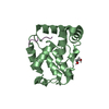
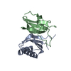
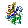
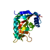
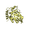
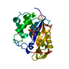
- PDB-1j1f: Crystal structure of the RNase MC1 mutant N71T in complex with 5'-GMP -
+ Open data

- Basic information
| Entry | Database: PDB / ID: 1j1f | ||||||
|---|---|---|---|---|---|---|---|
| Title | Crystal structure of the RNase MC1 mutant N71T in complex with 5'-GMP | ||||||
 Components Components | RIBONUCLEASE MC1 | ||||||
 Keywords Keywords | HYDROLASE / Nucleic acid / RNA | ||||||
| Function / homology |  Function and homology information Function and homology informationribonuclease T2 / ribonuclease T2 activity / RNA catabolic process / RNA binding / extracellular region Similarity search - Function | ||||||
| Biological species |  Momordica charantia (bitter melon) Momordica charantia (bitter melon) | ||||||
| Method |  X-RAY DIFFRACTION / SYNCHROTRON / MOLECULAR REPLACEMENT / Resolution: 1.6 Å X-RAY DIFFRACTION / SYNCHROTRON / MOLECULAR REPLACEMENT / Resolution: 1.6 Å | ||||||
 Authors Authors | Numata, T. / Suzuki, A. / Kakuta, Y. / Kimura, K. / Yao, M. / Tanaka, I. / Yoshida, Y. / Ueda, T. / Kimura, M. | ||||||
 Citation Citation | Journal: Biochemistry / Year: 2003 Title: Crystal Structures of the Ribonuclease MC1 Mutants N71T and N71S in Complex with 5'-GMP: Structural Basis for Alterations in Substrate Specificity Authors: Numata, T. / Suzuki, A. / Kakuta, Y. / Kimura, K. / Yao, M. / Tanaka, I. / Yoshida, Y. / Ueda, T. / Kimura, M. #1: Journal: BIOCHIM.BIOPHYS.ACTA / Year: 1999Title: Crystal structure of a ribonuclease from the seeds of bitter gourd (Momordica charantia) at 1.75 A resolution Authors: Nakagawa, A. / Tanaka, I. / Sakai, R. / Nakashima, T. / Funatsu, G. / Kimura, M. #2: Journal: Biochem.Biophys.Res.Commun. / Year: 2000Title: Crystal structures of the ribonuclease MC1 from bitter gourd seeds, complexed with 2'-UMP or 3'-UMP, reveal structural basis for uridine specificity Authors: Suzuki, A. / Yao, M. / Tanaka, I. / Numata, T. / Kikukawa, S. / Yamasaki, N. / Kimura, M. #3: Journal: BIOCHEMISTRY / Year: 2001Title: Amino acid residues in ribonuclease MC1 from bitter gourd seeds which are essential for uridine specificity Authors: Numata, T. / Suzuki, A. / Yao, M. / Tanaka, I. / Kimura, M. | ||||||
| History |
|
- Structure visualization
| Structure viewer | Molecule: MolmilJmol/JSmol |
|---|
- Downloads & links
-Download
| PDBx/mmCIF format | 1j1f.cif.gz | 55 KB | Display | PDBx/mmCIF format |
|---|---|---|---|---|
| PDB format | pdb1j1f.ent.gz | 38.7 KB | Display | PDB format |
| PDBx/mmJSON format | 1j1f.json.gz | Tree view | PDBx/mmJSON format | |
| Others |  Other downloads Other downloads |
-Validation report
| Summary document | 1j1f_validation.pdf.gz | 778.3 KB | Display | wwPDB validaton report |
|---|---|---|---|---|
| Full document | 1j1f_full_validation.pdf.gz | 779.5 KB | Display | |
| Data in XML | 1j1f_validation.xml.gz | 10.8 KB | Display | |
| Data in CIF | 1j1f_validation.cif.gz | 14.8 KB | Display | |
| Arichive directory | https://data.pdbj.org/pub/pdb/validation_reports/j1/1j1fftp://data.pdbj.org/pub/pdb/validation_reports/j1/1j1f | HTTPS FTP |
-Related structure data
| Related structure data |  1j1gC  1ucgC  1bk7S C: citing same article ( S: Starting model for refinement |
|---|---|
| Similar structure data |

-Links
 PDBj
PDBj

- Assembly
| Deposited unit | 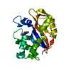
| ||||||||
|---|---|---|---|---|---|---|---|---|---|
| 1 |
| ||||||||
| Unit cell |
|
-Components
| #1: Protein | Mass: 21344.109 Da / Num. of mol.: 1 / Mutation: N71T Source method: isolated from a genetically manipulated source Source: (gene. exp.) Momordica charantia (bitter melon) / Plasmid: pPIC9K / Production host:  Pichia pastoris (fungus) / References: UniProt: P23540, EC: 3.1.27.1 Pichia pastoris (fungus) / References: UniProt: P23540, EC: 3.1.27.1 |
|---|---|
| #2: Chemical | ChemComp-5GP /   Mass: 363.221 Da / Num. of mol.: 1 / Source method: obtained synthetically / Formula: C10H14N5O8P Mass: 363.221 Da / Num. of mol.: 1 / Source method: obtained synthetically / Formula: C10H14N5O8P |
| #3: Water | ChemComp-HOH /  Mass: 18.015 Da / Num. of mol.: 136 / Source method: isolated from a natural source / Formula: H2O Mass: 18.015 Da / Num. of mol.: 136 / Source method: isolated from a natural source / Formula: H2O |
| Has protein modification | Y |
-Experimental details
-Experiment
| Experiment | Method: X-RAY DIFFRACTION / Number of used crystals: 1 |
|---|
- Sample preparation
| Crystal | Density Matthews: 2.06 Å3/Da / Density % sol: 39.69 % |
|---|---|
| Crystal grow | Temperature: 293 K / Method: vapor diffusion, hanging drop / pH: 5.6 Details: 0.2M ammonium acetate, 0.1M tri-sodium citrate, 27.5% PEG8000, pH 5.6, VAPOR DIFFUSION, HANGING DROP, temperature 293K |
-Data collection
| Diffraction | Mean temperature: 100 K |
|---|---|
| Diffraction source | Source: SYNCHROTRON / Site: SPring-8  / Beamline: BL44B2 / Wavelength: 1 Å / Beamline: BL44B2 / Wavelength: 1 Å |
| Detector | Type: MARRESEARCH / Detector: CCD / Date: Mar 1, 2002 / Details: mirrors |
| Radiation | Monochromator: Si 111 CHANNEL / Protocol: SINGLE WAVELENGTH / Monochromatic (M) / Laue (L): M / Scattering type: x-ray |
| Radiation wavelength | Wavelength: 1 Å / Relative weight: 1 |
| Reflection | Resolution: 1.5→50 Å / Num. all: 31008 / Num. obs: 31008 / % possible obs: 98.5 % / Observed criterion σ(F): 0 / Observed criterion σ(I): 0 / Redundancy: 5.1 % / Biso Wilson estimate: 15.8 Å2 / Rmerge(I) obs: 0.087 / Net I/σ(I): 15.7 |
| Reflection shell | Resolution: 1.5→1.55 Å / Redundancy: 4.5 % / Rmerge(I) obs: 0.201 / Mean I/σ(I) obs: 3 / Num. unique all: 2890 / % possible all: 92.9 |
- Processing
| Software |
| ||||||||||||||||||||||||||||||||||||||||||||||||||||||||||||
|---|---|---|---|---|---|---|---|---|---|---|---|---|---|---|---|---|---|---|---|---|---|---|---|---|---|---|---|---|---|---|---|---|---|---|---|---|---|---|---|---|---|---|---|---|---|---|---|---|---|---|---|---|---|---|---|---|---|---|---|---|---|
| Refinement | Method to determine structure: MOLECULAR REPLACEMENT Starting model: PDB ENTRY 1BK7 Resolution: 1.6→19.04 Å / Rfactor Rfree error: 0.007 / Isotropic thermal model: RESTRAINED / Cross valid method: THROUGHOUT / σ(F): 0 / σ(I): 0 / Stereochemistry target values: Engh & Huber
| ||||||||||||||||||||||||||||||||||||||||||||||||||||||||||||
| Solvent computation | Solvent model: FLAT MODEL / Bsol: 36.1703 Å2 / ksol: 0.367496 e/Å3 | ||||||||||||||||||||||||||||||||||||||||||||||||||||||||||||
| Displacement parameters | Biso mean: 17.9 Å2
| ||||||||||||||||||||||||||||||||||||||||||||||||||||||||||||
| Refine analyze |
| ||||||||||||||||||||||||||||||||||||||||||||||||||||||||||||
| Refinement step | Cycle: LAST / Resolution: 1.6→19.04 Å
| ||||||||||||||||||||||||||||||||||||||||||||||||||||||||||||
| Refine LS restraints |
| ||||||||||||||||||||||||||||||||||||||||||||||||||||||||||||
| LS refinement shell | Resolution: 1.6→1.7 Å / Rfactor Rfree error: 0.021 / Total num. of bins used: 6
| ||||||||||||||||||||||||||||||||||||||||||||||||||||||||||||
| Xplor file |
|
