 Movie
Movie Controller

[English] 日本語
 Yorodumi
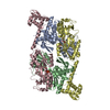
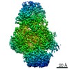
- PDB-1iz1: CRYSTAL STRUCTURE OF CBNR, A LYSR FAMILY TRANSCRIPTIONAL REGULATOR -
+ Open data

- Basic information
| Entry | Database: PDB / ID: 1iz1 | ||||||
|---|---|---|---|---|---|---|---|
| Title | CRYSTAL STRUCTURE OF CBNR, A LYSR FAMILY TRANSCRIPTIONAL REGULATOR | ||||||
 Components Components | LysR-type regulatory protein | ||||||
 Keywords Keywords | DNA BINDING PROTEIN / LONG ALPHA HELIX CONNECTING DNA BINDING AND REGULATORY DOMAINS | ||||||
| Function / homology |  Function and homology information Function and homology informationprotein-DNA complex / DNA-binding transcription factor activity / DNA binding Similarity search - Function | ||||||
| Biological species |  Cupriavidus necator (bacteria) Cupriavidus necator (bacteria) | ||||||
| Method |  X-RAY DIFFRACTION / SYNCHROTRON / MOLECULAR REPLACEMENT / Resolution: 2.5 Å X-RAY DIFFRACTION / SYNCHROTRON / MOLECULAR REPLACEMENT / Resolution: 2.5 Å | ||||||
 Authors Authors | Muraoka, S. / Okumura, R. / Ogawa, N. / Miyashita, K. / Senda, T. | ||||||
 Citation Citation | Journal: J.Mol.Biol. / Year: 2003 Title: Crystal Structure of a Full-length LysR-type Transcriptional Regulator, CbnR: Unusual Combination of Two Subunit Forms and Molecular Bases for Causing and Changing DNA Bend Authors: Muraoka, S. / Okumura, R. / Ogawa, N. / Nonaka, T. / Miyashita, K. / Senda, T. | ||||||
| History |
|
- Structure visualization
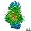
| Structure viewer | Molecule: MolmilJmol/JSmol |
|---|
- Downloads & links
-Download
| PDBx/mmCIF format | 1iz1.cif.gz | 224.2 KB | Display | PDBx/mmCIF format |
|---|---|---|---|---|
| PDB format | pdb1iz1.ent.gz | 183.6 KB | Display | PDB format |
| PDBx/mmJSON format | 1iz1.json.gz | Tree view | PDBx/mmJSON format | |
| Others |  Other downloads Other downloads |
-Validation report
| Arichive directory | https://data.pdbj.org/pub/pdb/validation_reports/iz/1iz1ftp://data.pdbj.org/pub/pdb/validation_reports/iz/1iz1 | HTTPS FTP |
|---|
-Related structure data
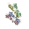
| Related structure data |  1ixcSC S: Starting model for refinement C: citing same article ( |
|---|---|
| Similar structure data |


-Links
 PDBj
PDBj- Assembly
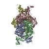
| Deposited unit | 
| ||||||||
|---|---|---|---|---|---|---|---|---|---|
| 1 |
| ||||||||
| Unit cell |
|
-Components
| #1: Protein | Mass: 32085.215 Da / Num. of mol.: 4 Source method: isolated from a genetically manipulated source Source: (gene. exp.) Cupriavidus necator (bacteria) / Plasmid: pET / Species (production host): Escherichia coli / Production host: #2: Water | ChemComp-HOH / |  Mass: 18.015 Da / Num. of mol.: 115 / Source method: isolated from a natural source / Formula: H2O Mass: 18.015 Da / Num. of mol.: 115 / Source method: isolated from a natural source / Formula: H2O |
|---|
-Experimental details
-Experiment
| Experiment | Method: X-RAY DIFFRACTION / Number of used crystals: 1 |
|---|
- Sample preparation
| Crystal | Density Matthews: 2.66 Å3/Da / Density % sol: 53.41 % |
|---|---|
| Crystal grow | Temperature: 293 K / Method: vapor diffusion, hanging drop / pH: 8 Details: Magnesium sulfate, Sodium chloride, Tris-HCl, pH 8.0, VAPOR DIFFUSION, HANGING DROP, temperature 293K |
| Crystal grow | *PLUS Details: Muraoka, S., (2003) Protein Pept. Lett., 10, 325. |
-Data collection
| Diffraction | Mean temperature: 100 K |
|---|---|
| Diffraction source | Source: SYNCHROTRON / Site: SPring-8  / Beamline: BL40B2 / Wavelength: 0.9797 Å / Beamline: BL40B2 / Wavelength: 0.9797 Å |
| Detector | Type: ADSC QUANTUM 4 / Detector: CCD / Date: Mar 22, 2001 |
| Radiation | Monochromator: FIXED-EXIT DOUBLE CRYSTAL MONOCHROMATOR SI(111) Protocol: SINGLE WAVELENGTH / Monochromatic (M) / Laue (L): M / Scattering type: x-ray |
| Radiation wavelength | Wavelength: 0.9797 Å / Relative weight: 1 |
| Reflection | Resolution: 2.5→43.5 Å / Num. all: 46950 / Num. obs: 46835 / % possible obs: 98.1 % / Observed criterion σ(F): 0 / Observed criterion σ(I): 0 / Redundancy: 4.5 % / Biso Wilson estimate: 42.9 Å2 / Rmerge(I) obs: 0.093 / Rsym value: 0.093 / Net I/σ(I): 6 |
| Reflection shell | Resolution: 2.5→2.64 Å / Redundancy: 3.1 % / Rmerge(I) obs: 0.389 / Mean I/σ(I) obs: 1.8 / Num. unique all: 284 / Rsym value: 0.389 / % possible all: 73.9 |
| Reflection | *PLUS Num. obs: 44422 / % possible obs: 97.8 % |
- Processing
| Software |
| ||||||||||||||||||||||||||||||||||||||||||||||||||||||||||||||||||||||||||||||||||||||||||||||||||||||||||||||||||||||||||||||||||
|---|---|---|---|---|---|---|---|---|---|---|---|---|---|---|---|---|---|---|---|---|---|---|---|---|---|---|---|---|---|---|---|---|---|---|---|---|---|---|---|---|---|---|---|---|---|---|---|---|---|---|---|---|---|---|---|---|---|---|---|---|---|---|---|---|---|---|---|---|---|---|---|---|---|---|---|---|---|---|---|---|---|---|---|---|---|---|---|---|---|---|---|---|---|---|---|---|---|---|---|---|---|---|---|---|---|---|---|---|---|---|---|---|---|---|---|---|---|---|---|---|---|---|---|---|---|---|---|---|---|---|---|
| Refinement | Method to determine structure: MOLECULAR REPLACEMENT Starting model: PDB ENTRY 1IXC Resolution: 2.5→50 Å / Cor.coef. Fo:Fc: 0.917 / Cor.coef. Fo:Fc free: 0.857 / SU B: 12.429 / SU ML: 0.267 / Cross valid method: THROUGHOUT / σ(F): 0 / ESU R: 0.607 / ESU R Free: 0.332 / Stereochemistry target values: MAXIMUM LIKELIHOOD
| ||||||||||||||||||||||||||||||||||||||||||||||||||||||||||||||||||||||||||||||||||||||||||||||||||||||||||||||||||||||||||||||||||
| Solvent computation | Ion probe radii: 0.8 Å / Shrinkage radii: 0.8 Å / VDW probe radii: 1.4 Å / Solvent model: BABINET MODEL WITH MASK | ||||||||||||||||||||||||||||||||||||||||||||||||||||||||||||||||||||||||||||||||||||||||||||||||||||||||||||||||||||||||||||||||||
| Displacement parameters | Biso mean: 30.719 Å2
| ||||||||||||||||||||||||||||||||||||||||||||||||||||||||||||||||||||||||||||||||||||||||||||||||||||||||||||||||||||||||||||||||||
| Refine analyze | Luzzati coordinate error free: 0.332 Å | ||||||||||||||||||||||||||||||||||||||||||||||||||||||||||||||||||||||||||||||||||||||||||||||||||||||||||||||||||||||||||||||||||
| Refinement step | Cycle: LAST / Resolution: 2.5→50 Å
| ||||||||||||||||||||||||||||||||||||||||||||||||||||||||||||||||||||||||||||||||||||||||||||||||||||||||||||||||||||||||||||||||||
| Refine LS restraints |
| ||||||||||||||||||||||||||||||||||||||||||||||||||||||||||||||||||||||||||||||||||||||||||||||||||||||||||||||||||||||||||||||||||
| LS refinement shell | Resolution: 2.5→2.565 Å / Total num. of bins used: 20 /
| ||||||||||||||||||||||||||||||||||||||||||||||||||||||||||||||||||||||||||||||||||||||||||||||||||||||||||||||||||||||||||||||||||
| Refinement | *PLUS Lowest resolution: 50 Å / Rfactor Rfree: 0.286 / Rfactor Rwork: 0.222 | ||||||||||||||||||||||||||||||||||||||||||||||||||||||||||||||||||||||||||||||||||||||||||||||||||||||||||||||||||||||||||||||||||
| Solvent computation | *PLUS | ||||||||||||||||||||||||||||||||||||||||||||||||||||||||||||||||||||||||||||||||||||||||||||||||||||||||||||||||||||||||||||||||||
| Displacement parameters | *PLUS | ||||||||||||||||||||||||||||||||||||||||||||||||||||||||||||||||||||||||||||||||||||||||||||||||||||||||||||||||||||||||||||||||||
| Refine LS restraints | *PLUS
|
