 Movie
Movie Controller


+ Open data

- Basic information
| Entry | Database: PDB / ID: 1ixs | ||||||
|---|---|---|---|---|---|---|---|
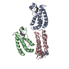
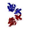
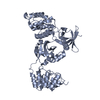
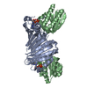
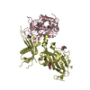
| Title | Structure of RuvB complexed with RuvA domain III | ||||||
 Components Components |
| ||||||
 Keywords Keywords | HYDROLASE / heterodimeric protein complex / AAA-ATPase domain / complex with nucleotide | ||||||
| Function / homology |  Function and homology information Function and homology informationHolliday junction helicase complex / Holliday junction resolvase complex / four-way junction helicase activity / four-way junction DNA binding / DNA recombination / DNA helicase / DNA repair / ATP hydrolysis activity / ATP binding / cytoplasm Similarity search - Function | ||||||
| Biological species |   Thermus thermophilus (bacteria) Thermus thermophilus (bacteria) | ||||||
| Method |  X-RAY DIFFRACTION / SYNCHROTRON / MOLECULAR REPLACEMENT / Resolution: 3.2 Å X-RAY DIFFRACTION / SYNCHROTRON / MOLECULAR REPLACEMENT / Resolution: 3.2 Å | ||||||
 Authors Authors | Yamada, K. / Miyata, T. / Tsuchiya, D. / Oyama, T. / Fujiwara, Y. / Ohnishi, T. / Iwasaki, H. / Shinagawa, H. / Ariyoshi, M. / Mayanagi, K. / Morikawa, K. | ||||||
 Citation Citation | Journal: Mol.Cell / Year: 2002 Title: Crystal Structure of the RuvA-RuvB Complex: A Structural Basis for the Holliday Junction Migrating Motor Machinery Authors: Yamada, K. / Miyata, T. / Tsuchiya, D. / Oyama, T. / Fujiwara, Y. / Ohnishi, T. / Iwasaki, H. / Shinagawa, H. / Ariyoshi, M. / Mayanagi, K. / Morikawa, K. | ||||||
| History |
|
- Structure visualization
| Structure viewer | Molecule: MolmilJmol/JSmol |
|---|
- Downloads & links
-Download
| PDBx/mmCIF format | 1ixs.cif.gz | 77.8 KB | Display | PDBx/mmCIF format |
|---|---|---|---|---|
| PDB format | pdb1ixs.ent.gz | 58.8 KB | Display | PDB format |
| PDBx/mmJSON format | 1ixs.json.gz | Tree view | PDBx/mmJSON format | |
| Others |  Other downloads Other downloads |
-Validation report
| Summary document | 1ixs_validation.pdf.gz | 763.7 KB | Display | wwPDB validaton report |
|---|---|---|---|---|
| Full document | 1ixs_full_validation.pdf.gz | 784.7 KB | Display | |
| Data in XML | 1ixs_validation.xml.gz | 17.6 KB | Display | |
| Data in CIF | 1ixs_validation.cif.gz | 23 KB | Display | |
| Arichive directory | https://data.pdbj.org/pub/pdb/validation_reports/ix/1ixsftp://data.pdbj.org/pub/pdb/validation_reports/ix/1ixs | HTTPS FTP |
-Related structure data
| Related structure data |  1ixrC  1hqcS S: Starting model for refinement C: citing same article ( |
|---|---|
| Similar structure data |


-Links
 PDBj
PDBj


- Assembly
| Deposited unit | 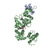
| ||||||||
|---|---|---|---|---|---|---|---|---|---|
| 1 |
| ||||||||
| Unit cell |
| ||||||||
| Details | The biological assembly is a heterodimer in the asymmetric unit. |
-Components
| #1: Protein | Mass: 6698.849 Da / Num. of mol.: 1 / Fragment: RuvA domain III Source method: isolated from a genetically manipulated source Source: (gene. exp.) Thermus thermophilus (bacteria) / Gene: ruva / Plasmid: pGEX / Species (production host): Escherichia coli / Production host: |
|---|---|
| #2: Protein | Mass: 35417.984 Da / Num. of mol.: 1 / Fragment: residues 1-318 Source method: isolated from a genetically manipulated source Source: (gene. exp.) Thermus thermophilus (bacteria) / Gene: ruvb / Plasmid: pET11a / Species (production host): Escherichia coli / Production host: |
| #3: Chemical | ChemComp-ANP /   Mass: 506.196 Da / Num. of mol.: 1 / Source method: obtained synthetically / Formula: C10H17N6O12P3 / Comment: AMP-PNP, energy-carrying molecule analogue*YM Mass: 506.196 Da / Num. of mol.: 1 / Source method: obtained synthetically / Formula: C10H17N6O12P3 / Comment: AMP-PNP, energy-carrying molecule analogue*YM |
-Experimental details
-Experiment
| Experiment | Method: X-RAY DIFFRACTION / Number of used crystals: 1 |
|---|
- Sample preparation
| Crystal | Density Matthews: 4.26 Å3/Da / Density % sol: 71.1 % | |||||||||||||||||||||||||||||||||||||||||||||||||||||||||||||||||||||||||||||
|---|---|---|---|---|---|---|---|---|---|---|---|---|---|---|---|---|---|---|---|---|---|---|---|---|---|---|---|---|---|---|---|---|---|---|---|---|---|---|---|---|---|---|---|---|---|---|---|---|---|---|---|---|---|---|---|---|---|---|---|---|---|---|---|---|---|---|---|---|---|---|---|---|---|---|---|---|---|---|
| Crystal grow | Temperature: 293 K / Method: vapor diffusion, hanging drop / pH: 8 Details: PEG 4000, pH 8.0, VAPOR DIFFUSION, HANGING DROP, temperature 293K | |||||||||||||||||||||||||||||||||||||||||||||||||||||||||||||||||||||||||||||
| Crystal grow | *PLUS Temperature: 20 ℃ | |||||||||||||||||||||||||||||||||||||||||||||||||||||||||||||||||||||||||||||
| Components of the solutions | *PLUS
|
-Data collection
| Diffraction | Mean temperature: 100 K |
|---|---|
| Diffraction source | Source: SYNCHROTRON / Site: SPring-8  / Beamline: BL24XU / Wavelength: 0.836 Å / Beamline: BL24XU / Wavelength: 0.836 Å |
| Detector | Type: RIGAKU RAXIS V / Detector: IMAGE PLATE / Date: Nov 13, 2001 |
| Radiation | Monochromator: DIAMOND / Protocol: SINGLE WAVELENGTH / Monochromatic (M) / Laue (L): M / Scattering type: x-ray |
| Radiation wavelength | Wavelength: 0.836 Å / Relative weight: 1 |
| Reflection | Resolution: 3.2→41 Å / Num. all: 235468 / Num. obs: 235468 / % possible obs: 99.5 % / Observed criterion σ(F): 0 / Observed criterion σ(I): 0 / Redundancy: 7.1 % / Rmerge(I) obs: 0.091 / Net I/σ(I): 6.8 |
| Reflection shell | Resolution: 3.2→3.37 Å / Redundancy: 7.3 % / Rmerge(I) obs: 0.324 / Mean I/σ(I) obs: 2.1 / % possible all: 99.5 |
| Reflection | *PLUS Lowest resolution: 41 Å / Num. obs: 12468 / Num. measured all: 235468 / Rmerge(I) obs: 0.091 |
| Reflection shell | *PLUS % possible obs: 99.5 % / Rmerge(I) obs: 0.324 |
- Processing
| Software |
| |||||||||||||||||||||||||
|---|---|---|---|---|---|---|---|---|---|---|---|---|---|---|---|---|---|---|---|---|---|---|---|---|---|---|
| Refinement | Method to determine structure: MOLECULAR REPLACEMENT Starting model: PDB ENTRY 1HQC Resolution: 3.2→40 Å / Isotropic thermal model: Isotropic / Cross valid method: THROUGHOUT / σ(F): 0 / Stereochemistry target values: Engh & Huber
| |||||||||||||||||||||||||
| Refine analyze |
| |||||||||||||||||||||||||
| Refinement step | Cycle: LAST / Resolution: 3.2→40 Å
| |||||||||||||||||||||||||
| Refine LS restraints |
| |||||||||||||||||||||||||
| LS refinement shell | Resolution: 3.2→3.31 Å
| |||||||||||||||||||||||||
| Refinement | *PLUS Lowest resolution: 40 Å / % reflection Rfree: 5 % / Rfactor Rfree: 0.293 / Rfactor Rwork: 0.23 | |||||||||||||||||||||||||
| Solvent computation | *PLUS | |||||||||||||||||||||||||
| Displacement parameters | *PLUS | |||||||||||||||||||||||||
| LS refinement shell | *PLUS Rfactor Rfree: 0.4718 / Rfactor Rwork: 0.35 |
