 Movie
Movie Controller

[English] 日本語
 Yorodumi
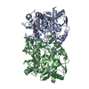
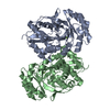
- PDB-1ixe: Crystal structure of citrate synthase from Thermus thermophilus HB8 -
+ Open data

- Basic information
| Entry | Database: PDB / ID: 1ixe | ||||||
|---|---|---|---|---|---|---|---|
| Title | Crystal structure of citrate synthase from Thermus thermophilus HB8 | ||||||
 Components Components | citrate synthase | ||||||
 Keywords Keywords | LYASE / enzyme-products complex / RIKEN Structural Genomics/Proteomics Initiative / RSGI / Structural Genomics | ||||||
| Function / homology |  Function and homology information Function and homology informationcitrate synthase activity / tricarboxylic acid cycle / carbohydrate metabolic process / cytosol Similarity search - Function | ||||||
| Biological species |   Thermus thermophilus (bacteria) Thermus thermophilus (bacteria) | ||||||
| Method |  X-RAY DIFFRACTION / SYNCHROTRON / MOLECULAR REPLACEMENT / Resolution: 2.3 Å X-RAY DIFFRACTION / SYNCHROTRON / MOLECULAR REPLACEMENT / Resolution: 2.3 Å | ||||||
 Authors Authors | Murakami, M. / Kanamori, E. / Kawaguchi, S. / Kuramitsu, S. / Kouyama, T. / RIKEN Structural Genomics/Proteomics Initiative (RSGI) | ||||||
 Citation Citation | Journal: Biophys Physicobio. / Year: 2015 Title: Structural comparison between the open and closed forms of citrate synthase from Thermus thermophilus HB8. Authors: Kanamori, E. / Kawaguchi, S. / Kuramitsu, S. / Kouyama, T. / Murakami, M. | ||||||
| History |
|
- Structure visualization
| Structure viewer | Molecule: MolmilJmol/JSmol |
|---|
- Downloads & links
-Download
| PDBx/mmCIF format | 1ixe.cif.gz | 308.2 KB | Display | PDBx/mmCIF format |
|---|---|---|---|---|
| PDB format | pdb1ixe.ent.gz | 250.9 KB | Display | PDB format |
| PDBx/mmJSON format | 1ixe.json.gz | Tree view | PDBx/mmJSON format | |
| Others |  Other downloads Other downloads |
-Validation report
| Arichive directory | https://data.pdbj.org/pub/pdb/validation_reports/ix/1ixeftp://data.pdbj.org/pub/pdb/validation_reports/ix/1ixe | HTTPS FTP |
|---|
-Related structure data
| Related structure data |  1iomSC S: Starting model for refinement C: citing same article ( |
|---|---|
| Similar structure data | |
| Other databases |

-Links
 PDBj
PDBj


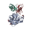
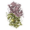
- Assembly
| Deposited unit | 
| ||||||||
|---|---|---|---|---|---|---|---|---|---|
| 1 | 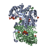
| ||||||||
| 2 | 
| ||||||||
| Unit cell |
| ||||||||
| Details | The biological assembly is a homodimer generated from the monomer in the asymmetric unit by the operations: y, x, -z. |
-Components
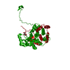
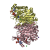
-Protein , 1 types, 4 molecules ABCD
| #1: Protein | Mass: 42362.441 Da / Num. of mol.: 4 Source method: isolated from a genetically manipulated source Source: (gene. exp.) Thermus thermophilus (bacteria) / Gene: HB8 / Plasmid: pET-3a / Production host: References: UniProt: Q9LCX9, UniProt: Q5SIM6*PLUS, EC: 4.1.3.7 |
|---|
-Non-polymers , 5 types, 539 molecules 


| #2: Chemical | ChemComp-SO4 /  Mass: 96.063 Da / Num. of mol.: 10 / Source method: obtained synthetically / Formula: SO4 Mass: 96.063 Da / Num. of mol.: 10 / Source method: obtained synthetically / Formula: SO4#3: Chemical | ChemComp-COA /  Mass: 767.534 Da / Num. of mol.: 4 / Source method: obtained synthetically / Formula: C21H36N7O16P3S Mass: 767.534 Da / Num. of mol.: 4 / Source method: obtained synthetically / Formula: C21H36N7O16P3S#4: Chemical | ChemComp-CIT /  Mass: 192.124 Da / Num. of mol.: 4 / Source method: obtained synthetically / Formula: C6H8O7 Mass: 192.124 Da / Num. of mol.: 4 / Source method: obtained synthetically / Formula: C6H8O7#5: Chemical | ChemComp-GOL /  Mass: 92.094 Da / Num. of mol.: 4 / Source method: obtained synthetically / Formula: C3H8O3 Mass: 92.094 Da / Num. of mol.: 4 / Source method: obtained synthetically / Formula: C3H8O3#6: Water | ChemComp-HOH / | Mass: 18.015 Da / Num. of mol.: 517 / Source method: isolated from a natural source / Formula: H2O |
|---|
-Experimental details
-Experiment
| Experiment | Method: X-RAY DIFFRACTION / Number of used crystals: 1 |
|---|
- Sample preparation
| Crystal | Density Matthews: 2.41 Å3/Da / Density % sol: 48.89 % |
|---|---|
| Crystal grow | Temperature: 293 K / Method: vapor diffusion, hanging drop / pH: 5.8 Details: lithium sulfate, sodium citrate, pH 5.8, VAPOR DIFFUSION, HANGING DROP, temperature 293K |
-Data collection
| Diffraction | Mean temperature: 100 K |
|---|---|
| Diffraction source | Source: SYNCHROTRON / Site: SPring-8  / Beamline: BL41XU / Wavelength: 1 Å / Beamline: BL41XU / Wavelength: 1 Å |
| Detector | Type: MARRESEARCH / Detector: CCD / Date: Jan 28, 2001 |
| Radiation | Monochromator: Si 111 / Protocol: SINGLE WAVELENGTH / Monochromatic (M) / Laue (L): M / Scattering type: x-ray |
| Radiation wavelength | Wavelength: 1 Å / Relative weight: 1 |
| Reflection | Resolution: 2.3→24.12 Å / Num. all: 73387 / Num. obs: 66309 / % possible obs: 90.7 % / Observed criterion σ(F): 0 / Observed criterion σ(I): 3 / Biso Wilson estimate: 18.1 Å2 / Rmerge(I) obs: 0.079 / Net I/σ(I): 7.8 |
| Reflection shell | Resolution: 2.3→2.44 Å / Redundancy: 8.2 % / Rmerge(I) obs: 0.217 / Mean I/σ(I) obs: 3.4 / Num. unique all: 8335 / Rsym value: 0.217 / % possible all: 76.7 |
- Processing
| Software |
| ||||||||||||||||||||||||||||||||||||
|---|---|---|---|---|---|---|---|---|---|---|---|---|---|---|---|---|---|---|---|---|---|---|---|---|---|---|---|---|---|---|---|---|---|---|---|---|---|
| Refinement | Method to determine structure: MOLECULAR REPLACEMENT Starting model: 1IOM Resolution: 2.3→14.79 Å / Rfactor Rfree error: 0.003 / Data cutoff high absF: 1489795.8 / Data cutoff low absF: 0 / Isotropic thermal model: RESTRAINED / Cross valid method: THROUGHOUT / σ(F): 0 / σ(I): 0 / Stereochemistry target values: Engh & Huber
| ||||||||||||||||||||||||||||||||||||
| Solvent computation | Solvent model: FLAT MODEL / Bsol: 30.8 Å2 / ksol: 0.372 e/Å3 | ||||||||||||||||||||||||||||||||||||
| Displacement parameters | Biso mean: 26.9 Å2
| ||||||||||||||||||||||||||||||||||||
| Refine analyze |
| ||||||||||||||||||||||||||||||||||||
| Refinement step | Cycle: LAST / Resolution: 2.3→14.79 Å
| ||||||||||||||||||||||||||||||||||||
| Refine LS restraints |
| ||||||||||||||||||||||||||||||||||||
| LS refinement shell | Resolution: 2.3→2.44 Å / Rfactor Rfree error: 0.008 / Total num. of bins used: 6
|
