 Movie
Movie Controller

[English] 日本語
 Yorodumi
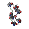
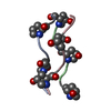
- PDB-1itt: Average Crystal Structure of (Pro-Pro-Gly)9 at 1.0 angstroms Reso... -
+ Open data

- Basic information
| Entry | Database: PDB / ID: 1itt | ||||||
|---|---|---|---|---|---|---|---|
| Title | Average Crystal Structure of (Pro-Pro-Gly)9 at 1.0 angstroms Resolution | ||||||
 Components Components | (COLLAGEN TRIPLE HELIX) x 3 | ||||||
 Keywords Keywords | STRUCTURAL PROTEIN / collagen / triple helix / Pro-Pro-Gly / single crystal | ||||||
| Method |  X-RAY DIFFRACTION / MOLECULAR REPLACEMENT / Resolution: 1 Å X-RAY DIFFRACTION / MOLECULAR REPLACEMENT / Resolution: 1 Å | ||||||
 Authors Authors | Hongo, C. / Nagarajan, V. / Noguchi, K. / Kamitori, S. / Okuyama, K. / Tanaka, Y. / Nishino, N. | ||||||
 Citation Citation | Journal: Plym.J. / Year: 2001 Title: Average Crystal Structure of (Pro-Pro-Gly)9 at 1.0 angstroms Resolution Authors: Hongo, C. / Nagarajan, V. / Noguchi, K. / Kamitori, S. / Okuyama, K. / Tanaka, Y. / Nishino, N. | ||||||
| History |
|
- Structure visualization
| Structure viewer | Molecule:  MolmilJmol/JSmol MolmilJmol/JSmol |
|---|
- Downloads & links
-Download
| PDBx/mmCIF format | 1itt.cif.gz | 10 KB | Display | PDBx/mmCIF format |
|---|---|---|---|---|
| PDB format | pdb1itt.ent.gz | 7 KB | Display | PDB format |
| PDBx/mmJSON format | 1itt.json.gz | Tree view | PDBx/mmJSON format | |
| Others |  Other downloads Other downloads |
-Validation report
| Summary document | 1itt_validation.pdf.gz | 341.2 KB | Display | wwPDB validaton report |
|---|---|---|---|---|
| Full document | 1itt_full_validation.pdf.gz | 341.2 KB | Display | |
| Data in XML | 1itt_validation.xml.gz | 1.6 KB | Display | |
| Data in CIF | 1itt_validation.cif.gz | 2.1 KB | Display | |
| Arichive directory | https://data.pdbj.org/pub/pdb/validation_reports/it/1ittftp://data.pdbj.org/pub/pdb/validation_reports/it/1itt | HTTPS FTP |
-Related structure data
| Similar structure data |
|---|


-Links
 PDBj
PDBj
- Assembly
| Deposited unit | 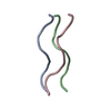
| ||||||||
|---|---|---|---|---|---|---|---|---|---|
| 1 |
| ||||||||
| Unit cell |
| ||||||||
| Details | THE ENTIRE 27 RESIDUE LONG PEPTIDE CAN BE GENERATED FROM THE SUBMITTED ASYMMETRIC UNIT BY APPLYING THE FOLLOWING TRANSLATIONS (USING FRACTIONAL COORDINATES): CHAIN A: TRANSLATE RESIDUES 2 - 7 BY (0 0 1), AND RESIDUES 1-7 BY (002), (003), (004) AND RESIDUE 1 BY (005). CHAIN B: TRANSLATE RESIDUES 33-37 BY (0 0 1), AND RESIDUES 31-37 BY (002), (003), (004) AND RESIDUE 31-32 BY (005). CHAIN C: TRANSLATE RESIDUES 64-67 BY (0 0 1), AND RESIDUES 61-67 BY (002), (003), (004). THIS WILL RESULT IN A MOLECULE WITH A TOTAL OF 81 RESIDUES, 27 IN EACH CHAIN. |
-Components
| #1: Protein/peptide | Mass: 577.630 Da / Num. of mol.: 1 / Source method: obtained synthetically Details: The peptide was chemically synthesized. The sequence of the peptide is naturally found in collagens. |
|---|---|
| #2: Protein/peptide | Mass: 617.693 Da / Num. of mol.: 1 / Source method: obtained synthetically Details: The peptide was chemically synthesized. The sequence of the peptide is naturally found in collagens. |
| #3: Protein/peptide | Mass: 617.693 Da / Num. of mol.: 1 / Source method: obtained synthetically Details: The peptide was chemically synthesized. The sequence of the peptide is naturally found in collagens. |
| #4: Water | ChemComp-HOH /  Mass: 18.015 Da / Num. of mol.: 34 / Source method: isolated from a natural source / Formula: H2O Mass: 18.015 Da / Num. of mol.: 34 / Source method: isolated from a natural source / Formula: H2O |
-Experimental details
-Experiment
| Experiment | Method: X-RAY DIFFRACTION / Number of used crystals: 1 |
|---|
- Sample preparation
| Crystal | Density Matthews: 1.96 Å3/Da / Density % sol: 37.13 % |
|---|---|
| Crystal grow | Temperature: 283 K / Method: vapor diffusion, hanging drop Details: PEG400, acetic acid, sodium azide, VAPOR DIFFUSION, HANGING DROP, temperature 283K |
-Data collection
| Diffraction | Mean temperature: 293 K |
|---|---|
| Diffraction source | Source: ROTATING ANODE / Type: RIGAKU RU200 / Wavelength: 1.5418 Å |
| Detector | Type: RIGAKU AFC-5R / Detector: DIFFRACTOMETER / Date: May 18, 1999 |
| Radiation | Monochromator: GRAPHITE / Protocol: SINGLE WAVELENGTH / Monochromatic (M) / Laue (L): M / Scattering type: x-ray |
| Radiation wavelength | Wavelength: 1.5418 Å / Relative weight: 1 |
| Reflection | Resolution: 1→8 Å / Num. all: 6129 / Num. obs: 2922 / Observed criterion σ(F): 1 |
- Processing
| Software |
| ||||||||||||||||||||
|---|---|---|---|---|---|---|---|---|---|---|---|---|---|---|---|---|---|---|---|---|---|
| Refinement | Method to determine structure: MOLECULAR REPLACEMENT Starting model: The structural model for collagen proposed by Okuyama (1981) Resolution: 1→8 Å / σ(F): 1 / Stereochemistry target values: Engh & Huber
| ||||||||||||||||||||
| Refinement step | Cycle: LAST / Resolution: 1→8 Å
| ||||||||||||||||||||
| Refine LS restraints |
|
