[English] 日本語
Yorodumi
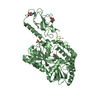
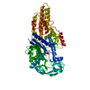
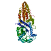
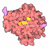
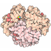
- PDB-1itb: TYPE-1 INTERLEUKIN-1 RECEPTOR COMPLEXED WITH INTERLEUKIN-1 BETA -

+
Open data


ID or keywords:

Loading...

-
Basic information

Entry
Database: PDB / ID: 1itb
TitleTYPE-1 INTERLEUKIN-1 RECEPTOR COMPLEXED WITH INTERLEUKIN-1 BETA
Components
  • INTERLEUKIN-1 BETA
  • TYPE 1 INTERLEUKIN-1 RECEPTOR
KeywordsCOMPLEX (IMMUNOGLOBULIN/RECEPTOR) / IMMUNOGLOBULIN FOLD / TRANSMEMBRANE / GLYCOPROTEIN / RECEPTOR / COMPLEX (IMMUNOGLOBULIN-RECEPTOR) / COMPLEX (IMMUNOGLOBULIN-RECEPTOR) complex
Function / homology
Function and homology information


interleukin-1, type I, activating receptor activity / interleukin-1 receptor activity / interleukin-1 binding / negative regulation of interleukin-1-mediated signaling pathway / positive regulation of T cell mediated immunity / negative regulation of adiponectin secretion / negative regulation of lipid metabolic process / smooth muscle adaptation / negative regulation of D-glucose transmembrane transport / positive regulation of lipid catabolic process ...interleukin-1, type I, activating receptor activity / interleukin-1 receptor activity / interleukin-1 binding / negative regulation of interleukin-1-mediated signaling pathway / positive regulation of T cell mediated immunity / negative regulation of adiponectin secretion / negative regulation of lipid metabolic process / smooth muscle adaptation / negative regulation of D-glucose transmembrane transport / positive regulation of lipid catabolic process / positive regulation of cell adhesion molecule production / regulation of nitric-oxide synthase activity / positive regulation of T-helper 1 cell cytokine production / hyaluronan biosynthetic process / positive regulation of complement activation / positive regulation of RNA biosynthetic process / cellular response to interleukin-17 / monocyte aggregation / positive regulation of tight junction disassembly / positive regulation of prostaglandin secretion / positive regulation of immature T cell proliferation in thymus / negative regulation of gap junction assembly / vascular endothelial growth factor production / positive regulation of prostaglandin biosynthetic process / positive regulation of fever generation / regulation of defense response to virus by host / fever generation / positive regulation of platelet-derived growth factor receptor signaling pathway / ADP-ribosyl cyclase/cyclic ADP-ribose hydrolase / CASP5-mediated substrate cleavage / CASP4-mediated substrate cleavage / NAD+ nucleosidase activity, cyclic ADP-ribose generating / CLEC7A/inflammasome pathway / regulation of establishment of endothelial barrier / Interleukin-1 processing / positive regulation of granulocyte macrophage colony-stimulating factor production / positive regulation of monocyte chemotactic protein-1 production / positive regulation of glial cell proliferation / positive regulation of macrophage derived foam cell differentiation / positive regulation of p38MAPK cascade / platelet-derived growth factor receptor binding / interleukin-1 receptor binding / negative regulation of synaptic transmission / positive regulation of vascular endothelial growth factor receptor signaling pathway / positive regulation of heterotypic cell-cell adhesion / positive regulation of membrane protein ectodomain proteolysis / regulation of canonical NF-kappaB signal transduction / interleukin-1-mediated signaling pathway / positive regulation of neuroinflammatory response / Interleukin-10 signaling / regulation of insulin secretion / positive regulation of cell division / negative regulation of extrinsic apoptotic signaling pathway in absence of ligand / positive regulation of vascular endothelial growth factor production / Pyroptosis / regulation of neurogenesis / regulation of ERK1 and ERK2 cascade / negative regulation of lipid catabolic process / positive regulation of epithelial to mesenchymal transition / embryo implantation / Purinergic signaling in leishmaniasis infection / negative regulation of MAPK cascade / positive regulation of protein export from nucleus / positive regulation of interleukin-2 production / negative regulation of insulin receptor signaling pathway / positive regulation of T cell proliferation / positive regulation of mitotic nuclear division / secretory granule / response to interleukin-1 / cytokine activity / positive regulation of interleukin-8 production / cellular response to xenobiotic stimulus / cellular response to mechanical stimulus / positive regulation of type II interferon production / negative regulation of inflammatory response / positive regulation of non-canonical NF-kappaB signal transduction / positive regulation of interleukin-6 production / negative regulation of neurogenesis / positive regulation of JNK cascade / integrin binding / cytokine-mediated signaling pathway / positive regulation of nitric oxide biosynthetic process / positive regulation of angiogenesis / positive regulation of inflammatory response / Interleukin-1 signaling / transmembrane signaling receptor activity / cell-cell signaling / cellular response to lipopolysaccharide / regulation of inflammatory response / response to lipopolysaccharide / protease binding / Interleukin-4 and Interleukin-13 signaling / positive regulation of phosphatidylinositol 3-kinase/protein kinase B signal transduction / Potential therapeutics for SARS / positive regulation of MAPK cascade / positive regulation of ERK1 and ERK2 cascade / positive regulation of canonical NF-kappaB signal transduction / cell surface receptor signaling pathway / lysosome / defense response to Gram-positive bacterium
Similarity search - Function
Interleukin-1 receptor type 1 / Interleukin-1 receptor type I/II / IL-1Ra-like, immunoglobulin domain / Immunoglobulin domain / Interleukin-1 receptor family / Interleukin-1 propeptide / Interleukin-1 propeptide / Interleukin-1 conserved site / Interleukin-1 signature. / Interleukin-1 homologues ...Interleukin-1 receptor type 1 / Interleukin-1 receptor type I/II / IL-1Ra-like, immunoglobulin domain / Immunoglobulin domain / Interleukin-1 receptor family / Interleukin-1 propeptide / Interleukin-1 propeptide / Interleukin-1 conserved site / Interleukin-1 signature. / Interleukin-1 homologues / Interleukin-1 family / Interleukin-1 / 18 / Cytokine IL1/FGF / TIR domain / Immunoglobulin domain / Toll - interleukin 1 - resistance / TIR domain profile. / Toll/interleukin-1 receptor homology (TIR) domain / Toll/interleukin-1 receptor homology (TIR) domain superfamily / Trefoil (Acidic Fibroblast Growth Factor, subunit A) - #50 / Trefoil (Acidic Fibroblast Growth Factor, subunit A) / Trefoil / Immunoglobulin subtype / Immunoglobulin / Ig-like domain profile. / Immunoglobulin-like domain / Immunoglobulin-like domain superfamily / Immunoglobulin-like fold / Immunoglobulins / Immunoglobulin-like / Sandwich / Mainly Beta
Similarity search - Domain/homology
Interleukin-1 beta / Interleukin-1 receptor type 1
Similarity search - Component
Biological speciesHomo sapiens (human)
MethodX-RAY DIFFRACTION / MIR / Resolution: 2.5 Å
AuthorsVigers, G.P.A. / Anderson, L.J. / Caffes, P. / Brandhuber, B.J.
CitationJournal: Nature / Year: 1997
Title: Crystal structure of the type-I interleukin-1 receptor complexed with interleukin-1beta.
Authors: Vigers, G.P. / Anderson, L.J. / Caffes, P. / Brandhuber, B.J.
History
DepositionJan 15, 1997Processing site: BNL
Revision 1.0Feb 4, 1998Provider: repository / Type: Initial release
Revision 1.1Mar 24, 2008Group: Version format compliance
Revision 1.2Jul 13, 2011Group: Version format compliance
Revision 1.3Oct 23, 2024Group: Data collection / Database references / Structure summary
Category: chem_comp_atom / chem_comp_bond ...chem_comp_atom / chem_comp_bond / database_2 / pdbx_entry_details / pdbx_modification_feature
Item: _database_2.pdbx_DOI / _database_2.pdbx_database_accession

-
Structure visualization

Structure viewerMolecule:
MolmilJmol/JSmol

Downloads & links

-
Assembly

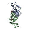
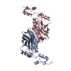
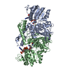
Deposited unit
A: INTERLEUKIN-1 BETA
B: TYPE 1 INTERLEUKIN-1 RECEPTOR


Theoretical massNumber of molelcules
Total (without water)53,6192
Polymers53,6192
Non-polymers00
Water43224
1


  • Idetical with deposited unit
  • defined by author
TypeNameSymmetry operationNumber
identity operation1_555x,y,z1
Unit cell
Length a, b, c (Å)146.952, 68.452, 65.867
Angle α, β, γ (deg.)90.00, 108.95, 90.00
Int Tables number5
Space group name H-MC121

-
Components

#1: Protein INTERLEUKIN-1 BETA


Mass: 17395.832 Da / Num. of mol.: 1
Source method: isolated from a genetically manipulated source
Source: (gene. exp.) Homo sapiens (human) / Cellular location: SECRETED / Production host: Escherichia coli (E. coli) / References: UniProt: P01584
#2: Protein TYPE 1 INTERLEUKIN-1 RECEPTOR


Mass: 36223.148 Da / Num. of mol.: 1 / Fragment: EXTRACELLULAR DOMAIN
Source method: isolated from a genetically manipulated source
Source: (gene. exp.) Homo sapiens (human) / Cellular location: CELL SURFACE / Production host: Escherichia coli (E. coli) / References: UniProt: P14778
#3: Water ChemComp-HOH / water


Mass: 18.015 Da / Num. of mol.: 24 / Source method: isolated from a natural source / Formula: H2O
Has protein modificationY

-
Experimental details

-
Experiment

ExperimentMethod: X-RAY DIFFRACTION / Number of used crystals: 2

-
Sample preparation

CrystalDensity Matthews: 2.92 Å3/Da / Density % sol: 52 %
Crystal growMethod: vapor diffusion, hanging drop / pH: 6
Details: PROTEIN WAS CRYSTALLIZED BY HANGING DROP DIFFUSION AGAINST 2.8M AMMONIUM SULFATE, 100MM MES PH 6.0, vapor diffusion - hanging drop
Crystal grow
*PLUS
Temperature: 0-4 ℃ / Method: vapor diffusion, hanging drop
Components of the solutions
*PLUS
IDConc.Common nameCrystal-IDSol-ID
11.8 Mammonium salfate1reservoir
2100 mMMES1reservoir

-
Data collection

DiffractionMean temperature: 100 K
Diffraction sourceSource: ROTATING ANODE / Type: RIGAKU RUH3R / Wavelength: 1.5418
DetectorType: RIGAKU / Detector: IMAGE PLATE / Date: Jul 1, 1995 / Details: COLLIMATOR
RadiationMonochromator: GRAPHITE(002) / Monochromatic (M) / Laue (L): M / Scattering type: x-ray
Radiation wavelengthWavelength: 1.5418 Å / Relative weight: 1
ReflectionResolution: 2.5→15 Å / Num. obs: 20723 / % possible obs: 96.4 % / Observed criterion σ(I): 2 / Redundancy: 6.4 % / Biso Wilson estimate: 37 Å2 / Rmerge(I) obs: 0.05 / Net I/σ(I): 28
Reflection shellResolution: 2.5→2.59 Å / Redundancy: 2.7 % / Rmerge(I) obs: 0.104 / Mean I/σ(I) obs: 10.9 / % possible all: 94.9

-
Processing

Software
NameClassification
DENZOdata reduction
SCALEPACKdata scaling
X-PLORmodel building
X-PLORrefinement
X-PLORphasing
RefinementMethod to determine structure: MIR / Resolution: 2.5→15 Å / Data cutoff high absF: 10000 / Data cutoff low absF: 0.1 / Isotropic thermal model: RESTRAINED / Cross valid method: THROUGHOUT / σ(F): 2
Details: BULK SOLVENT CORRECTION WITH SOLDENSITY = 0.40, SOLRAD = 0.25, B = 50
RfactorNum. reflection% reflectionSelection details
Rfree0.325 2061 9.6 %RANDOM
Rwork0.229 ---
obs0.229 20723 96.4 %-
Displacement parametersBiso mean: 53.7 Å2
Refine analyzeLuzzati sigma a obs: 0.44 Å
Refinement stepCycle: LAST / Resolution: 2.5→15 Å
ProteinNucleic acidLigandSolventTotal
Num. atoms3587 0 0 31 3618
Refine LS restraints
Refine-IDTypeDev idealDev ideal target
X-RAY DIFFRACTIONx_bond_d0.012
X-RAY DIFFRACTIONx_bond_d_na
X-RAY DIFFRACTIONx_bond_d_prot
X-RAY DIFFRACTIONx_angle_d
X-RAY DIFFRACTIONx_angle_d_na
X-RAY DIFFRACTIONx_angle_d_prot
X-RAY DIFFRACTIONx_angle_deg1.98
X-RAY DIFFRACTIONx_angle_deg_na
X-RAY DIFFRACTIONx_angle_deg_prot
X-RAY DIFFRACTIONx_dihedral_angle_d26.2
X-RAY DIFFRACTIONx_dihedral_angle_d_na
X-RAY DIFFRACTIONx_dihedral_angle_d_prot
X-RAY DIFFRACTIONx_improper_angle_d1.55
X-RAY DIFFRACTIONx_improper_angle_d_na
X-RAY DIFFRACTIONx_improper_angle_d_prot
X-RAY DIFFRACTIONx_mcbond_it1.5
X-RAY DIFFRACTIONx_mcangle_it2
X-RAY DIFFRACTIONx_scbond_it2
X-RAY DIFFRACTIONx_scangle_it2.5
LS refinement shellResolution: 2.5→2.56 Å / Total num. of bins used: 15
RfactorNum. reflection% reflection
Rfree0.5528 128 8.9 %
Rwork0.4308 1246 -
obs--95.5 %
Xplor file
Refine-IDSerial noParam fileTopol file
X-RAY DIFFRACTION1PARHCSDX.PROTOPHCSDX.PRO
X-RAY DIFFRACTION2PARAM19.SOLTOPH19.SOL
Software
*PLUS
Name: X-PLOR / Version: 3.1 / Classification: refinement
Refine LS restraints
*PLUS
Refine-IDTypeDev ideal
X-RAY DIFFRACTIONx_dihedral_angle_d
X-RAY DIFFRACTIONx_dihedral_angle_deg26.2
X-RAY DIFFRACTIONx_improper_angle_d
X-RAY DIFFRACTIONx_improper_angle_deg1.55

+
About Yorodumi

-
News

-
Feb 9, 2022. New format data for meta-information of EMDB entries

New format data for meta-information of EMDB entries

  • Version 3 of the EMDB header file is now the official format.
  • The previous official version 1.9 will be removed from the archive.

Related info.:EMDB header

External links:wwPDB to switch to version 3 of the EMDB data model

-
Aug 12, 2020. Covid-19 info

Covid-19 info

URL: https://pdbj.org/emnavi/covid19.php

New page: Covid-19 featured information page in EM Navigator.

Related info.:Covid-19 info / Mar 5, 2020. Novel coronavirus structure data

+
Mar 5, 2020. Novel coronavirus structure data

Novel coronavirus structure data

Related info.:Yorodumi Speices / Aug 12, 2020. Covid-19 info

External links:COVID-19 featured content - PDBj / Molecule of the Month (242):Coronavirus Proteases

+
Jan 31, 2019. EMDB accession codes are about to change! (news from PDBe EMDB page)

EMDB accession codes are about to change! (news from PDBe EMDB page)

  • The allocation of 4 digits for EMDB accession codes will soon come to an end. Whilst these codes will remain in use, new EMDB accession codes will include an additional digit and will expand incrementally as the available range of codes is exhausted. The current 4-digit format prefixed with “EMD-” (i.e. EMD-XXXX) will advance to a 5-digit format (i.e. EMD-XXXXX), and so on. It is currently estimated that the 4-digit codes will be depleted around Spring 2019, at which point the 5-digit format will come into force.
  • The EM Navigator/Yorodumi systems omit the EMD- prefix.

Related info.:Q: What is EMD? / ID/Accession-code notation in Yorodumi/EM Navigator

External links:EMDB Accession Codes are Changing Soon! / Contact to PDBj

+
Jul 12, 2017. Major update of PDB

Major update of PDB

  • wwPDB released updated PDB data conforming to the new PDBx/mmCIF dictionary.
  • This is a major update changing the version number from 4 to 5, and with Remediation, in which all the entries are updated.
  • In this update, many items about electron microscopy experimental information are reorganized (e.g. em_software).
  • Now, EM Navigator and Yorodumi are based on the updated data.

External links:wwPDB Remediation / Enriched Model Files Conforming to OneDep Data Standards Now Available in the PDB FTP Archive

-
Yorodumi

Thousand views of thousand structures

  • Yorodumi is a browser for structure data from EMDB, PDB, SASBDB, etc.
  • This page is also the successor to EM Navigator detail page, and also detail information page/front-end page for Omokage search.
  • The word "yorodu" (or yorozu) is an old Japanese word meaning "ten thousand". "mi" (miru) is to see.

Related info.:EMDB / PDB / SASBDB / Comparison of 3 databanks / Yorodumi Search / Aug 31, 2016. New EM Navigator & Yorodumi / Yorodumi Papers / Jmol/JSmol / Function and homology information / Changes in new EM Navigator and Yorodumi

Read more