 Movie
Movie Controller

[English] 日本語
 Yorodumi
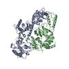
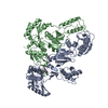
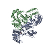
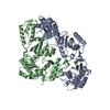
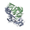
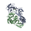
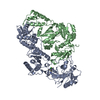
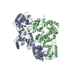
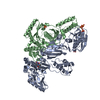
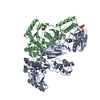
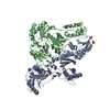
- PDB-1ikx: K103N Mutant HIV-1 Reverse Transcriptase in Complex with the Inhi... -
+ Open data

- Basic information
| Entry | Database: PDB / ID: 1ikx | ||||||
|---|---|---|---|---|---|---|---|
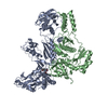
| Title | K103N Mutant HIV-1 Reverse Transcriptase in Complex with the Inhibitor PNU142721 | ||||||
 Components Components | (POL POLYPROTEIN) x 2 | ||||||
 Keywords Keywords | TRANSFERASE / Heterodimer / protein-inhibitor complex | ||||||
| Function / homology |  Function and homology information Function and homology informationRNA-directed DNA polymerase activity / HIV-1 retropepsin / symbiont-mediated activation of host apoptosis / retroviral ribonuclease H / exoribonuclease H / exoribonuclease H activity / host multivesicular body / DNA integration / viral genome integration into host DNA / RNA-directed DNA polymerase ...RNA-directed DNA polymerase activity / HIV-1 retropepsin / symbiont-mediated activation of host apoptosis / retroviral ribonuclease H / exoribonuclease H / exoribonuclease H activity / host multivesicular body / DNA integration / viral genome integration into host DNA / RNA-directed DNA polymerase / establishment of integrated proviral latency / telomerase activity / viral penetration into host nucleus / RNA stem-loop binding / RNA-DNA hybrid ribonuclease activity / Transferases; Transferring phosphorus-containing groups; Nucleotidyltransferases / host cell / viral nucleocapsid / DNA recombination / DNA-directed DNA polymerase / aspartic-type endopeptidase activity / Hydrolases; Acting on ester bonds / DNA-directed DNA polymerase activity / symbiont-mediated suppression of host gene expression / symbiont entry into host cell / viral translational frameshifting / lipid binding / host cell nucleus / host cell plasma membrane / virion membrane / structural molecule activity / proteolysis / DNA binding / zinc ion binding / membrane Similarity search - Function | ||||||
| Biological species |   Human immunodeficiency virus 1 Human immunodeficiency virus 1 | ||||||
| Method |  X-RAY DIFFRACTION / SYNCHROTRON / MOLECULAR REPLACEMENT / Resolution: 2.8 Å X-RAY DIFFRACTION / SYNCHROTRON / MOLECULAR REPLACEMENT / Resolution: 2.8 Å | ||||||
 Authors Authors | Lindberg, J. / Unge, T. | ||||||
 Citation Citation | Journal: Eur.J.Biochem. / Year: 2002 Title: Structural basis for the inhibitory efficacy of efavirenz (DMP-266), MSC194 and PNU142721 towards the HIV-1 RT K103N mutant. Authors: Lindberg, J. / Sigurdsson, S. / Lowgren, S. / Andersson, H.O. / Sahlberg, C. / Noreen, R. / Fridborg, K. / Zhang, H. / Unge, T. | ||||||
| History |
|
- Structure visualization
| Structure viewer | Molecule: MolmilJmol/JSmol |
|---|
- Downloads & links
-Download
| PDBx/mmCIF format | 1ikx.cif.gz | 213.2 KB | Display | PDBx/mmCIF format |
|---|---|---|---|---|
| PDB format | pdb1ikx.ent.gz | 168.9 KB | Display | PDB format |
| PDBx/mmJSON format | 1ikx.json.gz | Tree view | PDBx/mmJSON format | |
| Others |  Other downloads Other downloads |
-Validation report
| Arichive directory | https://data.pdbj.org/pub/pdb/validation_reports/ik/1ikxftp://data.pdbj.org/pub/pdb/validation_reports/ik/1ikx | HTTPS FTP |
|---|
-Related structure data
-Links
 PDBj
PDBj


- Assembly
| Deposited unit | 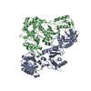
| ||||||||
|---|---|---|---|---|---|---|---|---|---|
| 1 |
| ||||||||
| 2 | 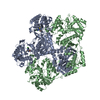
| ||||||||
| Unit cell |
| ||||||||
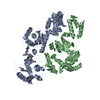
| Details | The biological assembly is a heterodimer |
-Components
| #1: Protein | Mass: 64500.965 Da / Num. of mol.: 1 / Mutation: K103N, E478Q Source method: isolated from a genetically manipulated source Source: (gene. exp.) Human immunodeficiency virus 1 / Genus: Lentivirus / Gene: BH10 / Plasmid: PETd / Species (production host): Escherichia coli / Production host:  |
|---|---|
| #2: Protein | Mass: 49912.344 Da / Num. of mol.: 1 / Mutation: K1103N Source method: isolated from a genetically manipulated source Source: (gene. exp.) Human immunodeficiency virus 1 / Genus: Lentivirus / Gene: BH10 / Plasmid: PETd / Species (production host): Escherichia coli / Production host: |
| #3: Chemical | ChemComp-PNU / 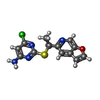  Mass: 306.771 Da / Num. of mol.: 1 / Source method: obtained synthetically / Formula: C13H11ClN4OS Mass: 306.771 Da / Num. of mol.: 1 / Source method: obtained synthetically / Formula: C13H11ClN4OS |
| #4: Water | ChemComp-HOH /  Mass: 18.015 Da / Num. of mol.: 61 / Source method: isolated from a natural source / Formula: H2O Mass: 18.015 Da / Num. of mol.: 61 / Source method: isolated from a natural source / Formula: H2O |
-Experimental details
-Experiment
| Experiment | Method: X-RAY DIFFRACTION / Number of used crystals: 5 |
|---|
- Sample preparation
| Crystal | Density Matthews: 3.21 Å3/Da / Density % sol: 61.71 % | |||||||||||||||||||||||||||||||||||||||||||||||||
|---|---|---|---|---|---|---|---|---|---|---|---|---|---|---|---|---|---|---|---|---|---|---|---|---|---|---|---|---|---|---|---|---|---|---|---|---|---|---|---|---|---|---|---|---|---|---|---|---|---|---|
| Crystal grow | Temperature: 295 K / Method: vapor diffusion, hanging drop / pH: 7.2 Details: Ammonium sulphate, pH 7.2, VAPOR DIFFUSION, HANGING DROP, temperature 295K | |||||||||||||||||||||||||||||||||||||||||||||||||
| Crystal grow | *PLUS Method: vapor diffusion | |||||||||||||||||||||||||||||||||||||||||||||||||
| Components of the solutions | *PLUS
|
-Data collection
| Diffraction | Mean temperature: 270 K |
|---|---|
| Diffraction source | Source: SYNCHROTRON / Site: MAX II  / Beamline: I711 / Wavelength: 1.0159 Å / Beamline: I711 / Wavelength: 1.0159 Å |
| Detector | Type: MARRESEARCH / Detector: IMAGE PLATE / Date: Feb 12, 2000 |
| Radiation | Monochromator: mirrors / Protocol: SINGLE WAVELENGTH / Monochromatic (M) / Laue (L): M / Scattering type: x-ray |
| Radiation wavelength | Wavelength: 1.0159 Å / Relative weight: 1 |
| Reflection | Resolution: 2.8→23.63 Å / Num. all: 40488 / Num. obs: 32921 / % possible obs: 81.3 % / Observed criterion σ(F): 2 / Observed criterion σ(I): 2 / Redundancy: 90.2 % / Biso Wilson estimate: 49.9 Å2 / Rmerge(I) obs: 0.075 / Net I/σ(I): 12.2 |
| Reflection shell | Resolution: 2.8→23.63 Å / Redundancy: 4.7 % / Rmerge(I) obs: 0.374 / % possible all: 92.8 |
| Reflection | *PLUS Num. measured all: 289120 |
| Reflection shell | *PLUS Highest resolution: 2.8 Å / Lowest resolution: 2.9 Å / % possible obs: 92.3 % / Num. unique obs: 3306 |
- Processing
| Software |
| ||||||||||||||||||||||||||||||||||||||||
|---|---|---|---|---|---|---|---|---|---|---|---|---|---|---|---|---|---|---|---|---|---|---|---|---|---|---|---|---|---|---|---|---|---|---|---|---|---|---|---|---|---|
| Refinement | Method to determine structure: MOLECULAR REPLACEMENT / Resolution: 2.8→23.63 Å / Rfactor Rfree error: 0.007 / Data cutoff high absF: 2936263.38 / Data cutoff low absF: 0 / Isotropic thermal model: RESTRAINED / Cross valid method: THROUGHOUT / σ(F): 2 / σ(I): 2 / Stereochemistry target values: Engh & Huber
| ||||||||||||||||||||||||||||||||||||||||
| Solvent computation | Solvent model: FLAT MODEL / Bsol: 33.92 Å2 / ksol: 0.291 e/Å3 | ||||||||||||||||||||||||||||||||||||||||
| Displacement parameters | Biso mean: 54.9 Å2
| ||||||||||||||||||||||||||||||||||||||||
| Refine analyze |
| ||||||||||||||||||||||||||||||||||||||||
| Refinement step | Cycle: LAST / Resolution: 2.8→23.63 Å
| ||||||||||||||||||||||||||||||||||||||||
| Refine LS restraints |
| ||||||||||||||||||||||||||||||||||||||||
| LS refinement shell | Resolution: 2.8→2.98 Å / Rfactor Rfree error: 0.02 / Total num. of bins used: 6
| ||||||||||||||||||||||||||||||||||||||||
| Xplor file |
| ||||||||||||||||||||||||||||||||||||||||
| Software | *PLUS Name: CNS / Version: 1 / Classification: refinement | ||||||||||||||||||||||||||||||||||||||||
| Refinement | *PLUS σ(F): 2 / % reflection Rfree: 5 % / Rfactor obs: 0.21 / Rfactor Rwork: 0.21 | ||||||||||||||||||||||||||||||||||||||||
| Solvent computation | *PLUS | ||||||||||||||||||||||||||||||||||||||||
| Displacement parameters | *PLUS Biso mean: 54.9 Å2 | ||||||||||||||||||||||||||||||||||||||||
| Refine LS restraints | *PLUS
| ||||||||||||||||||||||||||||||||||||||||
| LS refinement shell | *PLUS Rfactor Rfree: 0.337 / % reflection Rfree: 5 % / Rfactor Rwork: 0.295 |
