 Movie
Movie Controller


+ Open data

- Basic information
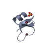
| Entry | Database: PDB / ID: 1iju | ||||||
|---|---|---|---|---|---|---|---|
| Title | HUMAN BETA-DEFENSIN-1 | ||||||
 Components Components | BETA-DEFENSIN 1 | ||||||
 Keywords Keywords | ANTIBIOTIC / DEFENSIN / HUMAN BETA-DEFENSIN-1 / BETA-DEFENSIN | ||||||
| Function / homology |  Function and homology information Function and homology informationpositive regulation of flagellated sperm motility involved in capacitation / CCR6 chemokine receptor binding / microvesicle / Beta defensins / Defensins / response to bacterium / innate immune response in mucosa / calcium-mediated signaling / Golgi lumen / chemotaxis ...positive regulation of flagellated sperm motility involved in capacitation / CCR6 chemokine receptor binding / microvesicle / Beta defensins / Defensins / response to bacterium / innate immune response in mucosa / calcium-mediated signaling / Golgi lumen / chemotaxis / antimicrobial humoral immune response mediated by antimicrobial peptide / antibacterial humoral response / sperm midpiece / defense response to Gram-negative bacterium / defense response to bacterium / defense response to Gram-positive bacterium / immune response / G protein-coupled receptor signaling pathway / innate immune response / : / extracellular exosome / extracellular region / membrane / identical protein binding Similarity search - Function | ||||||
| Method |  X-RAY DIFFRACTION / SYNCHROTRON / MOLECULAR REPLACEMENT / Resolution: 1.4 Å X-RAY DIFFRACTION / SYNCHROTRON / MOLECULAR REPLACEMENT / Resolution: 1.4 Å | ||||||
 Authors Authors | Hoover, D.M. / Lubkowski, J. | ||||||
 Citation Citation | Journal: J.Biol.Chem. / Year: 2001 Title: The structure of human beta-defensin-1: new insights into structural properties of beta-defensins. Authors: Hoover, D.M. / Chertov, O. / Lubkowski, J. | ||||||
| History |
|
- Structure visualization
| Structure viewer | Molecule: MolmilJmol/JSmol |
|---|
- Downloads & links
-Download
| PDBx/mmCIF format | 1iju.cif.gz | 50.3 KB | Display | PDBx/mmCIF format |
|---|---|---|---|---|
| PDB format | pdb1iju.ent.gz | 36.5 KB | Display | PDB format |
| PDBx/mmJSON format | 1iju.json.gz | Tree view | PDBx/mmJSON format | |
| Others |  Other downloads Other downloads |
-Validation report
| Arichive directory | https://data.pdbj.org/pub/pdb/validation_reports/ij/1ijuftp://data.pdbj.org/pub/pdb/validation_reports/ij/1iju | HTTPS FTP |
|---|
-Related structure data


-Links
 PDBj
PDBj

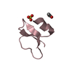
- Assembly
| Deposited unit | 
| ||||||||
|---|---|---|---|---|---|---|---|---|---|
| 1 | 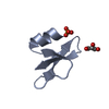
| ||||||||
| 2 | 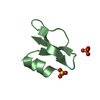
| ||||||||
| 3 | 
| ||||||||
| 4 | 
| ||||||||
| 5 | 
| ||||||||
| 6 | 
| ||||||||
| 7 | 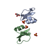
| ||||||||
| Unit cell |
|
-Components
| #1: Protein/peptide | Mass: 3940.598 Da / Num. of mol.: 4 / Source method: obtained synthetically Details: THIS PEPTIDE WAS CHEMICALLY SYNTHESIZED. THE SEQUENCE OF THIS PEPTIDE OCCURS NATURALLY IN HUMANS (HOMO SAPIENS). References: UniProt: P60022 #2: Chemical | ChemComp-SO4 /   Mass: 96.063 Da / Num. of mol.: 7 / Source method: obtained synthetically / Formula: SO4 Mass: 96.063 Da / Num. of mol.: 7 / Source method: obtained synthetically / Formula: SO4#3: Chemical |   Mass: 92.094 Da / Num. of mol.: 2 / Source method: obtained synthetically / Formula: C3H8O3 Mass: 92.094 Da / Num. of mol.: 2 / Source method: obtained synthetically / Formula: C3H8O3#4: Water | ChemComp-HOH / |  Mass: 18.015 Da / Num. of mol.: 271 / Source method: isolated from a natural source / Formula: H2O Mass: 18.015 Da / Num. of mol.: 271 / Source method: isolated from a natural source / Formula: H2OHas protein modification | Y | |
|---|
-Experimental details
-Experiment
| Experiment | Method: X-RAY DIFFRACTION / Number of used crystals: 1 |
|---|
- Sample preparation
| Crystal | Density Matthews: 1.77 Å3/Da / Density % sol: 30.4 % | ||||||||||||||||||||||||||||||||||||
|---|---|---|---|---|---|---|---|---|---|---|---|---|---|---|---|---|---|---|---|---|---|---|---|---|---|---|---|---|---|---|---|---|---|---|---|---|---|
| Crystal grow | Temperature: 285 K / Method: vapor diffusion, hanging drop / pH: 4.6 Details: PEG 4000, ammonium sulfate, sodium acetate, glycerol, pH 4.6, VAPOR DIFFUSION, HANGING DROP, temperature 285K | ||||||||||||||||||||||||||||||||||||
| Crystal grow | *PLUS | ||||||||||||||||||||||||||||||||||||
| Components of the solutions | *PLUS
|
-Data collection
| Diffraction | Mean temperature: 100 K | |||||||||
|---|---|---|---|---|---|---|---|---|---|---|
| Diffraction source | Source: SYNCHROTRON / Site: NSLS  / Beamline: X9B / Wavelength: 0.98 / Wavelength: 0.979 Å / Beamline: X9B / Wavelength: 0.98 / Wavelength: 0.979 Å | |||||||||
| Detector | Type: ADSC / Detector: CCD / Date: Mar 4, 2001 / Details: MIRRORS | |||||||||
| Radiation | Monochromator: SI CRYSTAL / Protocol: SINGLE WAVELENGTH / Monochromatic (M) / Laue (L): M / Scattering type: x-ray | |||||||||
| Radiation wavelength |
| |||||||||
| Reflection | Resolution: 1.4→20 Å / Num. all: 87587 / Num. obs: 26325 / % possible obs: 95.5 % / Observed criterion σ(F): 0 / Observed criterion σ(I): 0 / Redundancy: 3.3 % / Biso Wilson estimate: 15 Å2 / Rmerge(I) obs: 0.042 / Net I/σ(I): 27.2 | |||||||||
| Reflection shell | Resolution: 1.4→1.45 Å / Redundancy: 1.8 % / Rmerge(I) obs: 0.242 / Mean I/σ(I) obs: 2.5 / % possible all: 64.1 | |||||||||
| Reflection | *PLUS Highest resolution: 1.4 Å / Num. measured all: 87587 | |||||||||
| Reflection shell | *PLUS % possible obs: 64.1 % |
- Processing
| Software |
| |||||||||||||||||||||||||||||||||
|---|---|---|---|---|---|---|---|---|---|---|---|---|---|---|---|---|---|---|---|---|---|---|---|---|---|---|---|---|---|---|---|---|---|---|
| Refinement | Method to determine structure: MOLECULAR REPLACEMENT Starting model: HUMAN BETA-DEFENSIN-1 Resolution: 1.4→20 Å / Num. parameters: 6199 / Num. restraintsaints: 5551 / Cross valid method: FREE R / σ(F): 0 / σ(I): 0 / Stereochemistry target values: ENGH AND HUBER
| |||||||||||||||||||||||||||||||||
| Refine analyze | Num. disordered residues: 1 / Occupancy sum hydrogen: 1040 / Occupancy sum non hydrogen: 1350 | |||||||||||||||||||||||||||||||||
| Refinement step | Cycle: LAST / Resolution: 1.4→20 Å
| |||||||||||||||||||||||||||||||||
| Refine LS restraints |
| |||||||||||||||||||||||||||||||||
| Software | *PLUS Name: SHELXL-97 / Classification: refinement | |||||||||||||||||||||||||||||||||
| Refinement | *PLUS σ(F): 0 / % reflection Rfree: 4.4 % | |||||||||||||||||||||||||||||||||
| Solvent computation | *PLUS | |||||||||||||||||||||||||||||||||
| Displacement parameters | *PLUS | |||||||||||||||||||||||||||||||||
| Refine LS restraints | *PLUS
|
