 Movie
Movie Controller


+ Open data

- Basic information
| Entry | Database: PDB / ID: 1iir | ||||||
|---|---|---|---|---|---|---|---|
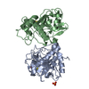
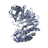
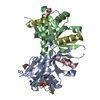
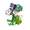
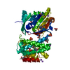
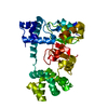
| Title | Crystal Structure of UDP-glucosyltransferase GtfB | ||||||
 Components Components | glycosyltransferase GtfB | ||||||
 Keywords Keywords | TRANSFERASE / glycosyltransferase / rossmann fold | ||||||
| Function / homology |  Function and homology information Function and homology informationvancomycin aglycone glucosyltransferase / vancomycin biosynthetic process / UDP-glycosyltransferase activity / hexosyltransferase activity / carbohydrate metabolic process Similarity search - Function | ||||||
| Biological species |  Amycolatopsis orientalis (bacteria) Amycolatopsis orientalis (bacteria) | ||||||
| Method |  X-RAY DIFFRACTION / SYNCHROTRON / MIR / Resolution: 1.8 Å X-RAY DIFFRACTION / SYNCHROTRON / MIR / Resolution: 1.8 Å | ||||||
 Authors Authors | Mulichak, A.M. / Losey, H.C. / Walsh, C.T. / Garavito, R.M. | ||||||
 Citation Citation | Journal: Structure / Year: 2001 Title: Structure of the UDP-glucosyltransferase GtfB that modifies the heptapeptide aglycone in the biosynthesis of vancomycin group antibiotics. Authors: Mulichak, A.M. / Losey, H.C. / Walsh, C.T. / Garavito, R.M. | ||||||
| History |
|
- Structure visualization
| Structure viewer | Molecule: MolmilJmol/JSmol |
|---|
- Downloads & links
-Download
| PDBx/mmCIF format | 1iir.cif.gz | 90 KB | Display | PDBx/mmCIF format |
|---|---|---|---|---|
| PDB format | pdb1iir.ent.gz | 66.9 KB | Display | PDB format |
| PDBx/mmJSON format | 1iir.json.gz | Tree view | PDBx/mmJSON format | |
| Others |  Other downloads Other downloads |
-Validation report
| Arichive directory | https://data.pdbj.org/pub/pdb/validation_reports/ii/1iirftp://data.pdbj.org/pub/pdb/validation_reports/ii/1iir | HTTPS FTP |
|---|
-Related structure data
| Similar structure data |
|---|
-Links
 PDBj
PDBj- Assembly
| Deposited unit | 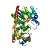
| |||||||||||||||||||||
|---|---|---|---|---|---|---|---|---|---|---|---|---|---|---|---|---|---|---|---|---|---|---|
| 1 |
| |||||||||||||||||||||
| Unit cell |
| |||||||||||||||||||||
| Components on special symmetry positions |
| |||||||||||||||||||||
| Details | Enzyme is a monomer. |
-Components
| #1: Protein | Mass: 43777.434 Da / Num. of mol.: 1 Source method: isolated from a genetically manipulated source Source: (gene. exp.) Amycolatopsis orientalis (bacteria) / Strain: A82846 / Plasmid: pET22b / Species (production host): Escherichia coli / Production host: References: UniProt: P96559, Transferases; Glycosyltransferases; Hexosyltransferases | ||
|---|---|---|---|
| #2: Chemical | ChemComp-SO4 /   Mass: 96.063 Da / Num. of mol.: 1 / Source method: obtained synthetically / Formula: SO4 Mass: 96.063 Da / Num. of mol.: 1 / Source method: obtained synthetically / Formula: SO4 | ||
| #3: Chemical |   Mass: 24.305 Da / Num. of mol.: 2 / Source method: obtained synthetically / Formula: Mg Mass: 24.305 Da / Num. of mol.: 2 / Source method: obtained synthetically / Formula: Mg#4: Water | ChemComp-HOH / |  Mass: 18.015 Da / Num. of mol.: 293 / Source method: isolated from a natural source / Formula: H2O Mass: 18.015 Da / Num. of mol.: 293 / Source method: isolated from a natural source / Formula: H2O |
-Experimental details
-Experiment
| Experiment | Method: X-RAY DIFFRACTION / Number of used crystals: 1 |
|---|
- Sample preparation
| Crystal | Density Matthews: 2.48 Å3/Da / Density % sol: 50.39 % | ||||||||||||||||||||||||||||||
|---|---|---|---|---|---|---|---|---|---|---|---|---|---|---|---|---|---|---|---|---|---|---|---|---|---|---|---|---|---|---|---|
| Crystal grow | Temperature: 298 K / Method: vapor diffusion, hanging drop / pH: 6.5 Details: magnesium sulfate, PEG 400, MES buffer, pH 6.5, VAPOR DIFFUSION, HANGING DROP, temperature 298K | ||||||||||||||||||||||||||||||
| Crystal grow | *PLUS | ||||||||||||||||||||||||||||||
| Components of the solutions | *PLUS
|
-Data collection
| Diffraction | Mean temperature: 100 K |
|---|---|
| Diffraction source | Source: SYNCHROTRON / Site: APS  / Beamline: 19-ID / Wavelength: 1.033 Å / Beamline: 19-ID / Wavelength: 1.033 Å |
| Detector | Type: CUSTOM-MADE / Detector: CCD / Date: Jun 8, 2000 |
| Radiation | Protocol: SINGLE WAVELENGTH / Monochromatic (M) / Laue (L): M / Scattering type: x-ray |
| Radiation wavelength | Wavelength: 1.033 Å / Relative weight: 1 |
| Reflection | Resolution: 1.8→30 Å / Num. all: 41142 / Num. obs: 41091 / % possible obs: 99.7 % / Observed criterion σ(F): 0 / Observed criterion σ(I): 0 / Redundancy: 6 % / Biso Wilson estimate: 18.8 Å2 / Rmerge(I) obs: 0.049 / Net I/σ(I): 26.8 |
| Reflection shell | Resolution: 1.8→1.86 Å / Redundancy: 6 % / Rmerge(I) obs: 0.281 / Mean I/σ(I) obs: 5 / Num. unique all: 4040 / % possible all: 100 |
- Processing
| Software |
| |||||||||||||||||||||||||
|---|---|---|---|---|---|---|---|---|---|---|---|---|---|---|---|---|---|---|---|---|---|---|---|---|---|---|
| Refinement | Method to determine structure: MIR / Resolution: 1.8→30 Å / σ(F): 1 / σ(I): 0 / Stereochemistry target values: Engh & Huber Details: Missing residues 56-62, 140-148, 246-248, 402-415 are disordered and unobserved in crystal structure. Also, for some residues side chain atoms are disordered and omitted from refined ...Details: Missing residues 56-62, 140-148, 246-248, 402-415 are disordered and unobserved in crystal structure. Also, for some residues side chain atoms are disordered and omitted from refined coordinates (C8, R11, E41, R63, E92, I149, D150, Q160, R273, D282, D283).
| |||||||||||||||||||||||||
| Refine analyze | Luzzati coordinate error obs: 0.22 Å / Luzzati sigma a obs: 0.13 Å | |||||||||||||||||||||||||
| Refinement step | Cycle: LAST / Resolution: 1.8→30 Å
| |||||||||||||||||||||||||
| Refine LS restraints |
| |||||||||||||||||||||||||
| LS refinement shell | Resolution: 1.8→1.86 Å
| |||||||||||||||||||||||||
| Software | *PLUS Name: CNS / Version: 0.9 / Classification: refinement | |||||||||||||||||||||||||
| Refinement | *PLUS Highest resolution: 1.8 Å / Lowest resolution: 30 Å / σ(F): 1 / % reflection Rfree: 7.2 % / Rfactor obs: 0.211 | |||||||||||||||||||||||||
| Solvent computation | *PLUS | |||||||||||||||||||||||||
| Displacement parameters | *PLUS | |||||||||||||||||||||||||
| Refine LS restraints | *PLUS
| |||||||||||||||||||||||||
| LS refinement shell | *PLUS Rfactor Rfree: 0.294 / Rfactor Rwork: 0.241 |
