 Movie
Movie Controller

[English] 日本語
 Yorodumi
- PDB-1iha: Structure of the Hybrid RNA/DNA R-GCUUCGGC-D[BR]U in Presence of ... -
+ Open data

- Basic information
| Entry | Database: PDB / ID: 1iha | ||||||||||||||||||
|---|---|---|---|---|---|---|---|---|---|---|---|---|---|---|---|---|---|---|---|
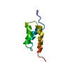
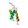
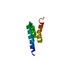
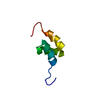
| Title | Structure of the Hybrid RNA/DNA R-GCUUCGGC-D[BR]U in Presence of RH(NH3)6+++ | ||||||||||||||||||
 Components Components | 5'-R(* Keywords KeywordsDNA/RNA / RNA/DNA HYBRID / BROMO URACIL / RHODIUM(III) HEXAMMINE / C-U G-U MISMATCH / DNA-RNA COMPLEX | Function / homology | RHODIUM HEXAMINE ION / DNA/RNA hybrid |  Function and homology information Function and homology informationMethod |  X-RAY DIFFRACTION / SYNCHROTRON / MOLECULAR REPLACEMENT / Resolution: 1.6 Å X-RAY DIFFRACTION / SYNCHROTRON / MOLECULAR REPLACEMENT / Resolution: 1.6 Å  Authors AuthorsCruse, W.B. / Saludjian, P. / Neuman, A. / Prange, T. |  CitationJournal: Acta Crystallogr.,Sect.D / Year: 2001 CitationJournal: Acta Crystallogr.,Sect.D / Year: 2001Title: Destabilizing effect of a fluorouracil extra base in a hybrid RNA duplex compared with bromo and chloro analogues. Authors: Cruse, W. / Saludjian, P. / Neuman, A. / Prange, T. #1: Journal: Proc.Natl.Acad.Sci.USA / Year: 1994Title: Structure of a Mispaired RNA Double Helix at 1.6 A Resolution and Implications for the Prediction of RNA Secondary Structure Authors: Cruse, W.B. / Saludjian, P. / Biala, E. / Strazewski, P. / Prange, T. / Kennard, O. History |
|
- Structure visualization
| Structure viewer | Molecule: MolmilJmol/JSmol |
|---|
- Downloads & links
-Download
| PDBx/mmCIF format | 1iha.cif.gz | 22.9 KB | Display | PDBx/mmCIF format |
|---|---|---|---|---|
| PDB format | pdb1iha.ent.gz | 14.6 KB | Display | PDB format |
| PDBx/mmJSON format | 1iha.json.gz | Tree view | PDBx/mmJSON format | |
| Others |  Other downloads Other downloads |
-Validation report
| Arichive directory | https://data.pdbj.org/pub/pdb/validation_reports/ih/1ihaftp://data.pdbj.org/pub/pdb/validation_reports/ih/1iha | HTTPS FTP |
|---|
-Related structure data
| Related structure data | 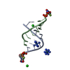 1icgC  1id9C 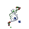 1idwC 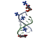 165dS S: Starting model for refinement C: citing same article ( |
|---|---|
| Similar structure data |


-Links
 PDBj
PDBj- Assembly
| Deposited unit | 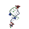
| ||||||||
|---|---|---|---|---|---|---|---|---|---|
| 1 |
| ||||||||
| Unit cell |
|
-Components
| #1: DNA/RNA hybrid | Mass: 2887.600 Da / Num. of mol.: 2 / Source method: obtained synthetically / Details: phosphoramidite method #2: Chemical |   Mass: 205.089 Da / Num. of mol.: 2 / Source method: obtained synthetically / Formula: H18N6Rh Mass: 205.089 Da / Num. of mol.: 2 / Source method: obtained synthetically / Formula: H18N6Rh#3: Chemical |   Mass: 35.453 Da / Num. of mol.: 2 / Source method: obtained synthetically / Formula: Cl Mass: 35.453 Da / Num. of mol.: 2 / Source method: obtained synthetically / Formula: Cl#4: Water | ChemComp-HOH / |  Mass: 18.015 Da / Num. of mol.: 61 / Source method: isolated from a natural source / Formula: H2O Mass: 18.015 Da / Num. of mol.: 61 / Source method: isolated from a natural source / Formula: H2O |
|---|
-Experimental details
-Experiment
| Experiment | Method: X-RAY DIFFRACTION / Number of used crystals: 1 |
|---|
- Sample preparation
| Crystal | Density Matthews: 2.18 Å3/Da / Density % sol: 43.7 % | ||||||||||||||||||||||||||||||||||||
|---|---|---|---|---|---|---|---|---|---|---|---|---|---|---|---|---|---|---|---|---|---|---|---|---|---|---|---|---|---|---|---|---|---|---|---|---|---|
| Crystal grow | Temperature: 277 K / Method: vapor diffusion, sitting drop / pH: 6.5 Details: MPD, Rhodium hexammine, lithium cacodylate, pH 6.5, VAPOR DIFFUSION, SITTING DROP, temperature 277K | ||||||||||||||||||||||||||||||||||||
| Components of the solutions |
| ||||||||||||||||||||||||||||||||||||
| Crystal grow | *PLUS | ||||||||||||||||||||||||||||||||||||
| Components of the solutions | *PLUS
|
-Data collection
| Diffraction | Mean temperature: 277 K |
|---|---|
| Diffraction source | Source: SYNCHROTRON / Site: LURE  / Beamline: DW32 / Beamline: DW32 |
| Detector | Type: MARRESEARCH / Detector: IMAGE PLATE / Date: Mar 5, 1995 Details: siliciu(111) curvated crystal plus multi-layer curvated mirror |
| Radiation | Monochromator: Si(111) / Protocol: SINGLE WAVELENGTH / Monochromatic (M) / Laue (L): M / Scattering type: x-ray |
| Radiation wavelength | Relative weight: 1 |
| Reflection | Resolution: 1.6→9.5 Å / Num. all: 6400 / Num. obs: 6312 / % possible obs: 98 % / Observed criterion σ(F): 3 / Observed criterion σ(I): 4 / Redundancy: 9 % / Biso Wilson estimate: 17 Å2 / Rmerge(I) obs: 0.045 / Rsym value: 0.043 / Net I/σ(I): 21 |
| Reflection shell | Resolution: 1.6→1.7 Å / Redundancy: 3 % / Rmerge(I) obs: 0.141 / Mean I/σ(I) obs: 3.8 / Num. unique all: 997 / Rsym value: 0.138 / % possible all: 89 |
| Reflection | *PLUS % possible obs: 96 % / Redundancy: 6.2 % / Num. measured all: 40122 |
- Processing
| Software |
| |||||||||||||||||||||||||||||||||||||||||||||||||||||||||||||||||||||||||||||||||||||||||||||||||||||||||||||||||||||||||
|---|---|---|---|---|---|---|---|---|---|---|---|---|---|---|---|---|---|---|---|---|---|---|---|---|---|---|---|---|---|---|---|---|---|---|---|---|---|---|---|---|---|---|---|---|---|---|---|---|---|---|---|---|---|---|---|---|---|---|---|---|---|---|---|---|---|---|---|---|---|---|---|---|---|---|---|---|---|---|---|---|---|---|---|---|---|---|---|---|---|---|---|---|---|---|---|---|---|---|---|---|---|---|---|---|---|---|---|---|---|---|---|---|---|---|---|---|---|---|---|---|---|---|
| Refinement | Method to determine structure: MOLECULAR REPLACEMENT Starting model: 165D from pdb Resolution: 1.6→1.75 Å / Isotropic thermal model: isotropic / Cross valid method: THROUGHOUT / σ(F): 2 / σ(I): 4 Stereochemistry target values: personal dictionary from small molecules Details: chain B (iso =11.8) is more agitated than chain A (iso =18.9)
Displacement parameters | Biso mean: 16.3 Å2 | Refinement step | Cycle: LAST / Resolution: 1.6→1.75 Å |
Refine LS restraints |
LS refinement shell |
Software | *PLUS Name: SHELXL-97 / Classification: refinementRefinement | *PLUS σ(F): 2 Solvent computation | *PLUS Displacement parameters | *PLUS Refine LS restraints | *PLUS
|
