 Movie
Movie Controller

[English] 日本語
 Yorodumi
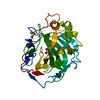
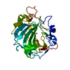
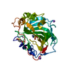
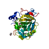
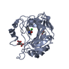
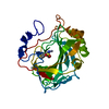
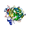
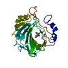
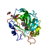
- PDB-1if8: Carbonic Anhydrase II Complexed With (S)-N-(3-Indol-1-yl-2-methyl... -
+ Open data

- Basic information
| Entry | Database: PDB / ID: 1if8 | ||||||
|---|---|---|---|---|---|---|---|
| Title | Carbonic Anhydrase II Complexed With (S)-N-(3-Indol-1-yl-2-methyl-propyl)-4-sulfamoyl-benzamide | ||||||
 Components Components | CARBONIC ANHYDRASE II | ||||||
 Keywords Keywords | LYASE / Carbonic Anhydrase II / (S)-N-(3-Indol-1-yl-2-methyl-propyl)-4-sulfamoyl-benzamide | ||||||
| Function / homology |  Function and homology information Function and homology informationpositive regulation of dipeptide transmembrane transport / : / secretion / regulation of monoatomic anion transport / cyanamide hydratase / cyanamide hydratase activity / arylesterase activity / regulation of chloride transport / Reversible hydration of carbon dioxide / positive regulation of synaptic transmission, GABAergic ...positive regulation of dipeptide transmembrane transport / : / secretion / regulation of monoatomic anion transport / cyanamide hydratase / cyanamide hydratase activity / arylesterase activity / regulation of chloride transport / Reversible hydration of carbon dioxide / positive regulation of synaptic transmission, GABAergic / morphogenesis of an epithelium / angiotensin-activated signaling pathway / Developmental Lineage of Pancreatic Ductal Cells / carbonic anhydrase / regulation of intracellular pH / carbonate dehydratase activity / carbon dioxide transport / neuron cellular homeostasis / Erythrocytes take up oxygen and release carbon dioxide / Erythrocytes take up carbon dioxide and release oxygen / apical part of cell / myelin sheath / extracellular exosome / zinc ion binding / plasma membrane / cytosol / cytoplasm Similarity search - Function | ||||||
| Biological species |  Homo sapiens (human) Homo sapiens (human) | ||||||
| Method |  X-RAY DIFFRACTION / FOURIER SYNTHESIS / Resolution: 1.94 Å X-RAY DIFFRACTION / FOURIER SYNTHESIS / Resolution: 1.94 Å | ||||||
 Authors Authors | Grzybowski, B.A. / Ishchenko, A.V. / Kim, C.-Y. / Topalov, G. / Chapman, R. / Christianson, D.W. / Whitesides, G.M. / Shakhnovich, E.I. | ||||||
 Citation Citation | Journal: Proc.Natl.Acad.Sci.USA / Year: 2002 Title: Combinatorial computational method gives new picomolar ligands for a known enzyme. Authors: Grzybowski, B.A. / Ishchenko, A.V. / Kim, C.Y. / Topalov, G. / Chapman, R. / Christianson, D.W. / Whitesides, G.M. / Shakhnovich, E.I. | ||||||
| History |
| ||||||
| Remark 999 | SEQUENCE THE RESIDUE NUMBERING IS NOT SEQUENTIAL. RESIDUE 125 IS COVALENTLY BOUND TO RESIDUE 127. |
- Structure visualization
| Structure viewer | Molecule: MolmilJmol/JSmol |
|---|
- Downloads & links
-Download
| PDBx/mmCIF format | 1if8.cif.gz | 67.3 KB | Display | PDBx/mmCIF format |
|---|---|---|---|---|
| PDB format | pdb1if8.ent.gz | 47.7 KB | Display | PDB format |
| PDBx/mmJSON format | 1if8.json.gz | Tree view | PDBx/mmJSON format | |
| Others |  Other downloads Other downloads |
-Validation report
| Arichive directory | https://data.pdbj.org/pub/pdb/validation_reports/if/1if8ftp://data.pdbj.org/pub/pdb/validation_reports/if/1if8 | HTTPS FTP |
|---|
-Related structure data
| Related structure data | 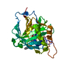 1if7C 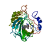 1if9C 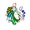 2cbaS C: citing same article ( S: Starting model for refinement |
|---|---|
| Similar structure data |


-Links
 PDBj
PDBj


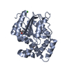
- Assembly
| Deposited unit | 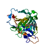
| ||||||||
|---|---|---|---|---|---|---|---|---|---|
| 1 |
| ||||||||
| Unit cell |
|
-Components
| #1: Protein | Mass: 29157.863 Da / Num. of mol.: 1 Source method: isolated from a genetically manipulated source Source: (gene. exp.) Homo sapiens (human) / Production host:  |
|---|---|
| #2: Chemical | ChemComp-ZN /   Mass: 65.409 Da / Num. of mol.: 1 / Source method: obtained synthetically / Formula: Zn Mass: 65.409 Da / Num. of mol.: 1 / Source method: obtained synthetically / Formula: Zn |
| #3: Chemical | ChemComp-HG /   Mass: 200.590 Da / Num. of mol.: 1 / Source method: obtained synthetically / Formula: Hg Mass: 200.590 Da / Num. of mol.: 1 / Source method: obtained synthetically / Formula: Hg |
| #4: Chemical | ChemComp-SBS / (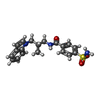  Mass: 371.453 Da / Num. of mol.: 1 / Source method: obtained synthetically / Formula: C19H21N3O3S Mass: 371.453 Da / Num. of mol.: 1 / Source method: obtained synthetically / Formula: C19H21N3O3S |
| #5: Water | ChemComp-HOH /  Mass: 18.015 Da / Num. of mol.: 67 / Source method: isolated from a natural source / Formula: H2O Mass: 18.015 Da / Num. of mol.: 67 / Source method: isolated from a natural source / Formula: H2O |
-Experimental details
-Experiment
| Experiment | Method: X-RAY DIFFRACTION / Number of used crystals: 1 |
|---|
- Sample preparation
| Crystal | Density Matthews: 2.24 Å3/Da / Density % sol: 45.05 % | ||||||||||||||||||||||||||||||||||||
|---|---|---|---|---|---|---|---|---|---|---|---|---|---|---|---|---|---|---|---|---|---|---|---|---|---|---|---|---|---|---|---|---|---|---|---|---|---|
| Crystal grow | Temperature: 277 K / Method: vapor diffusion, hanging drop / pH: 8 Details: methyl mercuric acetate, tris-sulfate, ammonium sulfate, pH 8.0, VAPOR DIFFUSION, HANGING DROP, temperature 277K | ||||||||||||||||||||||||||||||||||||
| Crystal grow | *PLUS Temperature: 4 ℃Details: drop consists of equal amounts of protein and precipitant solutions | ||||||||||||||||||||||||||||||||||||
| Components of the solutions | *PLUS
|
-Data collection
| Diffraction | Mean temperature: 298 K |
|---|---|
| Diffraction source | Source: ROTATING ANODE / Type: RIGAKU RU200 / Wavelength: 1.54 Å |
| Detector | Type: RIGAKU RAXIS IIC / Detector: IMAGE PLATE / Date: Mar 29, 2000 |
| Radiation | Monochromator: YALE MIRRORS / Protocol: SINGLE WAVELENGTH / Monochromatic (M) / Laue (L): M / Scattering type: x-ray |
| Radiation wavelength | Wavelength: 1.54 Å / Relative weight: 1 |
| Reflection | Resolution: 1.94→20 Å / Num. all: 18866 / Num. obs: 16222 / Observed criterion σ(F): 2 / Redundancy: 4 % / Biso Wilson estimate: 17.8 Å2 / Rmerge(I) obs: 0.122 / Net I/σ(I): 7.4 |
| Reflection | *PLUS Num. obs: 18866 / % possible obs: 94 % / Num. measured all: 76137 |
- Processing
| Software |
| |||||||||||||||
|---|---|---|---|---|---|---|---|---|---|---|---|---|---|---|---|---|
| Refinement | Method to determine structure: FOURIER SYNTHESIS Starting model: PDB ENTRY 2CBA Resolution: 1.94→20 Å / σ(F): 2
| |||||||||||||||
| Refinement step | Cycle: LAST / Resolution: 1.94→20 Å
| |||||||||||||||
| Refine LS restraints |
| |||||||||||||||
| Software | *PLUS Name: X-PLOR / Classification: refinement | |||||||||||||||
| Refine LS restraints | *PLUS
|
