 Movie
Movie Controller

[English] 日本語
 Yorodumi
- PDB-1icw: INTERLEUKIN-8, MUTANT WITH GLU 38 REPLACED BY CYS AND CYS 50 REPL... -
+ Open data

- Basic information
| Entry | Database: PDB / ID: 1icw | ||||||
|---|---|---|---|---|---|---|---|
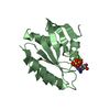
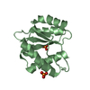
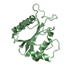
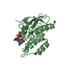
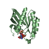
| Title | INTERLEUKIN-8, MUTANT WITH GLU 38 REPLACED BY CYS AND CYS 50 REPLACED BY ALA | ||||||
 Components Components | INTERLEUKIN-8 | ||||||
 Keywords Keywords | CYTOKINE / CHEMOKINE | ||||||
| Function / homology |  Function and homology information Function and homology informationregulation of single stranded viral RNA replication via double stranded DNA intermediate / regulation of entry of bacterium into host cell / interleukin-8 receptor binding / negative regulation of cell adhesion molecule production / ATF4 activates genes in response to endoplasmic reticulum stress / CXCR chemokine receptor binding / embryonic digestive tract development / neutrophil activation / induction of positive chemotaxis / positive regulation of neutrophil chemotaxis ...regulation of single stranded viral RNA replication via double stranded DNA intermediate / regulation of entry of bacterium into host cell / interleukin-8 receptor binding / negative regulation of cell adhesion molecule production / ATF4 activates genes in response to endoplasmic reticulum stress / CXCR chemokine receptor binding / embryonic digestive tract development / neutrophil activation / induction of positive chemotaxis / positive regulation of neutrophil chemotaxis / chemokine activity / Chemokine receptors bind chemokines / negative regulation of G protein-coupled receptor signaling pathway / Interleukin-10 signaling / cellular response to interleukin-1 / regulation of cell adhesion / neutrophil chemotaxis / cellular response to fibroblast growth factor stimulus / response to endoplasmic reticulum stress / Peptide ligand-binding receptors / response to molecule of bacterial origin / calcium-mediated signaling / receptor internalization / cellular response to tumor necrosis factor / chemotaxis / positive regulation of angiogenesis / antimicrobial humoral immune response mediated by antimicrobial peptide / heparin binding / cellular response to lipopolysaccharide / Senescence-Associated Secretory Phenotype (SASP) / Interleukin-4 and Interleukin-13 signaling / angiogenesis / G alpha (i) signalling events / intracellular signal transduction / G protein-coupled receptor signaling pathway / inflammatory response / negative regulation of cell population proliferation / negative regulation of gene expression / positive regulation of gene expression / signal transduction / : / extracellular region / membrane Similarity search - Function | ||||||
| Biological species |  Homo sapiens (human) Homo sapiens (human) | ||||||
| Method |  X-RAY DIFFRACTION / Resolution: 2.01 Å X-RAY DIFFRACTION / Resolution: 2.01 Å | ||||||
 Authors Authors | Eigenbrot, C. / Lowman, H.B. / Chee, L. / Artis, D.R. | ||||||
 Citation Citation | Journal: Proteins / Year: 1997 Title: Structural change and receptor binding in a chemokine mutant with a rearranged disulfide: X-ray structure of E38C/C50AIL-8 at 2 A resolution. Authors: Eigenbrot, C. / Lowman, H.B. / Chee, L. / Artis, D.R. #1: Journal: Proc.Natl.Acad.Sci.USA / Year: 1991Title: Crystal Structure of Interleukin 8: Symbiosis of NMR and Crystallography Authors: Baldwin, E.T. / Weber, I.T. / Charles, R.St. / Xuan, J.C. / Appella, E. / Yamada, M. / Matsushima, K. / Edwards, B.F. / Clore, G.M. / Gronenborn, A.M. / al., et | ||||||
| History |
|
- Structure visualization
| Structure viewer | Molecule: MolmilJmol/JSmol |
|---|
- Downloads & links
-Download
| PDBx/mmCIF format | 1icw.cif.gz | 40.1 KB | Display | PDBx/mmCIF format |
|---|---|---|---|---|
| PDB format | pdb1icw.ent.gz | 28.2 KB | Display | PDB format |
| PDBx/mmJSON format | 1icw.json.gz | Tree view | PDBx/mmJSON format | |
| Others |  Other downloads Other downloads |
-Validation report
| Arichive directory | https://data.pdbj.org/pub/pdb/validation_reports/ic/1icwftp://data.pdbj.org/pub/pdb/validation_reports/ic/1icw | HTTPS FTP |
|---|
-Related structure data
| Similar structure data |
|---|


-Links
 PDBj
PDBj


- Assembly
| Deposited unit | 
| ||||||||
|---|---|---|---|---|---|---|---|---|---|
| 1 |
| ||||||||
| Unit cell |
|
-Components
| #1: Protein | Mass: 8343.771 Da / Num. of mol.: 2 / Mutation: E38C, C50A Source method: isolated from a genetically manipulated source Source: (gene. exp.) Homo sapiens (human) / Production host:  #2: Water | ChemComp-HOH / |  Mass: 18.015 Da / Num. of mol.: 52 / Source method: isolated from a natural source / Formula: H2O Mass: 18.015 Da / Num. of mol.: 52 / Source method: isolated from a natural source / Formula: H2OHas protein modification | Y | |
|---|
-Experimental details
-Experiment
| Experiment | Method: X-RAY DIFFRACTION / Number of used crystals: 1 |
|---|
- Sample preparation
| Crystal | Density Matthews: 2.38 Å3/Da / Density % sol: 42 % | ||||||||||||||||||||||||||||||||||||
|---|---|---|---|---|---|---|---|---|---|---|---|---|---|---|---|---|---|---|---|---|---|---|---|---|---|---|---|---|---|---|---|---|---|---|---|---|---|
| Crystal grow | *PLUS pH: 6.2 / Method: vapor diffusion, sitting dropDetails: protein solution is mixed in a 5:3 ratio with well solution | ||||||||||||||||||||||||||||||||||||
| Components of the solutions | *PLUS
|
-Data collection
| Diffraction | Mean temperature: 291 K |
|---|---|
| Diffraction source | Source: ROTATING ANODE / Wavelength: 1.5418 |
| Detector | Type: MARRESEARCH / Detector: IMAGE PLATE / Date: Jun 28, 1995 |
| Radiation | Monochromator: GRAPHITE(002) / Monochromatic (M) / Laue (L): M / Scattering type: x-ray |
| Radiation wavelength | Wavelength: 1.5418 Å / Relative weight: 1 |
| Reflection | Resolution: 2.01→15 Å / Num. obs: 10544 / % possible obs: 96 % / Redundancy: 5 % / Rmerge(I) obs: 0.043 / Net I/σ(I): 27 |
| Reflection shell | Resolution: 2.01→2.09 Å / Redundancy: 4 % / % possible all: 78 |
| Reflection shell | *PLUS % possible obs: 78 % |
- Processing
| Software |
| ||||||||||||||||||||||||||||||||||||||||||||||||||||||||||||
|---|---|---|---|---|---|---|---|---|---|---|---|---|---|---|---|---|---|---|---|---|---|---|---|---|---|---|---|---|---|---|---|---|---|---|---|---|---|---|---|---|---|---|---|---|---|---|---|---|---|---|---|---|---|---|---|---|---|---|---|---|---|
| Refinement | Resolution: 2.01→10 Å / σ(F): 2
| ||||||||||||||||||||||||||||||||||||||||||||||||||||||||||||
| Displacement parameters | Biso mean: 32 Å2 | ||||||||||||||||||||||||||||||||||||||||||||||||||||||||||||
| Refinement step | Cycle: LAST / Resolution: 2.01→10 Å
| ||||||||||||||||||||||||||||||||||||||||||||||||||||||||||||
| Refine LS restraints |
| ||||||||||||||||||||||||||||||||||||||||||||||||||||||||||||
| LS refinement shell | Resolution: 2.01→2.09 Å /
| ||||||||||||||||||||||||||||||||||||||||||||||||||||||||||||
| Software | *PLUS Name: X-PLOR / Classification: refinement | ||||||||||||||||||||||||||||||||||||||||||||||||||||||||||||
| Refinement | *PLUS | ||||||||||||||||||||||||||||||||||||||||||||||||||||||||||||
| Solvent computation | *PLUS | ||||||||||||||||||||||||||||||||||||||||||||||||||||||||||||
| Displacement parameters | *PLUS | ||||||||||||||||||||||||||||||||||||||||||||||||||||||||||||
| Refine LS restraints | *PLUS
|
