 Movie
Movie Controller

[English] 日本語
 Yorodumi
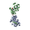
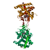
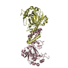
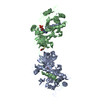
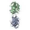
- PDB-1iaj: CRYSTAL STRUCTURE OF THE ATYPICAL PROTEIN KINASE DOMAIN OF A TRP ... -
+ Open data

- Basic information
| Entry | Database: PDB / ID: 1iaj | ||||||
|---|---|---|---|---|---|---|---|
| Title | CRYSTAL STRUCTURE OF THE ATYPICAL PROTEIN KINASE DOMAIN OF A TRP CA-CHANNEL, CHAK (APO) | ||||||
 Components Components | TRANSIENT RECEPTOR POTENTIAL-RELATED PROTEIN | ||||||
 Keywords Keywords | TRANSFERASE / alpha/beta / protein kinase like fold / ATP-grasp fold | ||||||
| Function / homology |  Function and homology information Function and homology informationcalcium-dependent cell-matrix adhesion / intracellular magnesium ion homeostasis / varicosity / magnesium ion transport / magnesium ion transmembrane transport / magnesium ion homeostasis / zinc ion transport / zinc ion transmembrane transporter activity / magnesium ion transmembrane transporter activity / TRP channels ...calcium-dependent cell-matrix adhesion / intracellular magnesium ion homeostasis / varicosity / magnesium ion transport / magnesium ion transmembrane transport / magnesium ion homeostasis / zinc ion transport / zinc ion transmembrane transporter activity / magnesium ion transmembrane transporter activity / TRP channels / actomyosin structure organization / myosin binding / necroptotic process / monoatomic cation transmembrane transport / monoatomic cation channel activity / ruffle / calcium ion transmembrane transport / calcium channel activity / memory / kinase activity / calcium ion transport / synaptic vesicle membrane / actin binding / cytoplasmic vesicle / protein homotetramerization / protein kinase activity / non-specific serine/threonine protein kinase / positive regulation of apoptotic process / protein serine kinase activity / protein serine/threonine kinase activity / neuronal cell body / ATP binding / metal ion binding / nucleus / plasma membrane Similarity search - Function | ||||||
| Biological species |  | ||||||
| Method |  X-RAY DIFFRACTION / SYNCHROTRON / MIR / Resolution: 2.8 Å X-RAY DIFFRACTION / SYNCHROTRON / MIR / Resolution: 2.8 Å | ||||||
 Authors Authors | Yamaguchi, H. / Matsushita, M. / Nairn, A.C. / Kuriyan, J. | ||||||
 Citation Citation | Journal: Mol.Cell / Year: 2001 Title: Crystal structure of the atypical protein kinase domain of a TRP channel with phosphotransferase activity. Authors: Yamaguchi, H. / Matsushita, M. / Nairn, A.C. / Kuriyan, J. | ||||||
| History |
|
- Structure visualization
| Structure viewer | Molecule: MolmilJmol/JSmol |
|---|
- Downloads & links
-Download
| PDBx/mmCIF format | 1iaj.cif.gz | 104.3 KB | Display | PDBx/mmCIF format |
|---|---|---|---|---|
| PDB format | pdb1iaj.ent.gz | 81.3 KB | Display | PDB format |
| PDBx/mmJSON format | 1iaj.json.gz | Tree view | PDBx/mmJSON format | |
| Others |  Other downloads Other downloads |
-Validation report
| Arichive directory | https://data.pdbj.org/pub/pdb/validation_reports/ia/1iajftp://data.pdbj.org/pub/pdb/validation_reports/ia/1iaj | HTTPS FTP |
|---|
-Related structure data


-Links
 PDBj
PDBj- Assembly
| Deposited unit | 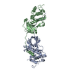
| ||||||||
|---|---|---|---|---|---|---|---|---|---|
| 1 |
| ||||||||
| Unit cell |
|
-Components
| #1: Protein | Mass: 32246.873 Da / Num. of mol.: 2 / Fragment: PROTEIN KINASE DOMAIN, RESIDUES 1549-1828 Source method: isolated from a genetically manipulated source Source: (gene. exp.)   Spodoptera frugiperda (fall armyworm) / Strain (production host): SF9 / References: UniProt: Q923J1, EC: 2.7.1.37 Spodoptera frugiperda (fall armyworm) / Strain (production host): SF9 / References: UniProt: Q923J1, EC: 2.7.1.37#2: Chemical |   Mass: 65.409 Da / Num. of mol.: 2 / Source method: obtained synthetically / Formula: Zn Mass: 65.409 Da / Num. of mol.: 2 / Source method: obtained synthetically / Formula: Zn |
|---|
-Experimental details
-Experiment
| Experiment | Method: X-RAY DIFFRACTION / Number of used crystals: 1 |
|---|
- Sample preparation
| Crystal | Density Matthews: 3.29 Å3/Da / Density % sol: 62.62 % | ||||||||||||||||||||||||||||||||||||||||
|---|---|---|---|---|---|---|---|---|---|---|---|---|---|---|---|---|---|---|---|---|---|---|---|---|---|---|---|---|---|---|---|---|---|---|---|---|---|---|---|---|---|
| Crystal grow | Temperature: 293 K / Method: vapor diffusion, hanging drop / pH: 7 Details: HEPES, PEG 4000, 2-propanol, pH 7.0, VAPOR DIFFUSION, HANGING DROP, temperature 293.0K | ||||||||||||||||||||||||||||||||||||||||
| Crystal grow | *PLUS Temperature: 20 ℃ | ||||||||||||||||||||||||||||||||||||||||
| Components of the solutions | *PLUS
|
-Data collection
| Diffraction | Mean temperature: 110 K |
|---|---|
| Diffraction source | Source: SYNCHROTRON / Site: NSLS  / Beamline: X25 / Wavelength: 1.1 Å / Beamline: X25 / Wavelength: 1.1 Å |
| Detector | Type: CUSTOM-MADE / Detector: CCD / Date: May 30, 1999 |
| Radiation | Monochromator: Si 111 / Protocol: SINGLE WAVELENGTH / Monochromatic (M) / Laue (L): M / Scattering type: x-ray |
| Radiation wavelength | Wavelength: 1.1 Å / Relative weight: 1 |
| Reflection | Resolution: 2.8→30 Å / Num. all: 21336 / Num. obs: 20775 / % possible obs: 97.4 % / Observed criterion σ(F): 0 / Observed criterion σ(I): 0 / Redundancy: 3.4 % / Biso Wilson estimate: 74.27 Å2 / Rmerge(I) obs: 0.049 / Rsym value: 4.9 / Net I/σ(I): 22.4 |
| Reflection shell | Resolution: 2.8→2.9 Å / Redundancy: 2.7 % / Rmerge(I) obs: 0.172 / Mean I/σ(I) obs: 3.7 / Num. unique all: 1979 / Rsym value: 17.2 / % possible all: 94.2 |
| Reflection shell | *PLUS % possible obs: 94.2 % |
- Processing
| Software |
| ||||||||||||||||||||
|---|---|---|---|---|---|---|---|---|---|---|---|---|---|---|---|---|---|---|---|---|---|
| Refinement | Method to determine structure: MIR / Resolution: 2.8→20 Å / Cross valid method: THROUGHOUT / σ(F): 2 / σ(I): 4 / Stereochemistry target values: Engh & Huber
| ||||||||||||||||||||
| Refinement step | Cycle: LAST / Resolution: 2.8→20 Å
| ||||||||||||||||||||
| Software | *PLUS Name: X-PLOR / Version: 3.851 / Classification: refinement | ||||||||||||||||||||
| Refinement | *PLUS σ(F): 2 | ||||||||||||||||||||
| Solvent computation | *PLUS | ||||||||||||||||||||
| Displacement parameters | *PLUS | ||||||||||||||||||||
| Refine LS restraints | *PLUS
|
