 Movie
Movie Controller


+ Open data

- Basic information
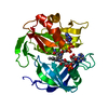
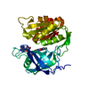
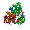
| Entry | Database: PDB / ID: 1i7p | ||||||
|---|---|---|---|---|---|---|---|
| Title | CRYSTAL STRUCTURE OF RAT B5R IN COMPLEX WITH FAD | ||||||
 Components Components | NADH-CYTOCHROME B5 REDUCTASE | ||||||
 Keywords Keywords | OXYGEN STORAGE/TRANSPORT / ELECTRON TRANSPORT / HEMOGLOBINEMIA / ERYTHROCYTE FUNCTION / FAD-BINDING / NADH-BINDING / OXYGEN STORAGE-TRANSPORT COMPLEX | ||||||
| Function / homology |  Function and homology information Function and homology informationPhase I - Functionalization of compounds / nitric-oxide synthase complex / Vitamin C (ascorbate) metabolism / cytochrome-b5 reductase activity, acting on NADH / cytochrome-b5 reductase / cytochrome-b5 reductase activity, acting on NAD(P)H / Neutrophil degranulation / cholesterol biosynthetic process / AMP binding / nitric oxide biosynthetic process ...Phase I - Functionalization of compounds / nitric-oxide synthase complex / Vitamin C (ascorbate) metabolism / cytochrome-b5 reductase activity, acting on NADH / cytochrome-b5 reductase / cytochrome-b5 reductase activity, acting on NAD(P)H / Neutrophil degranulation / cholesterol biosynthetic process / AMP binding / nitric oxide biosynthetic process / FAD binding / lipid droplet / ADP binding / mitochondrial membrane / NAD binding / flavin adenine dinucleotide binding / mitochondrial outer membrane / endoplasmic reticulum membrane / endoplasmic reticulum / mitochondrion / cytosol Similarity search - Function | ||||||
| Biological species |  | ||||||
| Method |  X-RAY DIFFRACTION / SYNCHROTRON / MOLECULAR REPLACEMENT / Resolution: 2 Å X-RAY DIFFRACTION / SYNCHROTRON / MOLECULAR REPLACEMENT / Resolution: 2 Å | ||||||
 Authors Authors | Bewley, M.C. / Marohnic, C.C. / Barber, M.J. | ||||||
 Citation Citation | Journal: Biochemistry / Year: 2001 Title: The structure and biochemistry of NADH-dependent cytochrome b5 reductase are now consistent. Authors: Bewley, M.C. / Marohnic, C.C. / Barber, M.J. | ||||||
| History |
|
- Structure visualization
| Structure viewer | Molecule: MolmilJmol/JSmol |
|---|
- Downloads & links
-Download
| PDBx/mmCIF format | 1i7p.cif.gz | 73 KB | Display | PDBx/mmCIF format |
|---|---|---|---|---|
| PDB format | pdb1i7p.ent.gz | 52.4 KB | Display | PDB format |
| PDBx/mmJSON format | 1i7p.json.gz | Tree view | PDBx/mmJSON format | |
| Others |  Other downloads Other downloads |
-Validation report
| Arichive directory | https://data.pdbj.org/pub/pdb/validation_reports/i7/1i7pftp://data.pdbj.org/pub/pdb/validation_reports/i7/1i7p | HTTPS FTP |
|---|
-Related structure data
| Related structure data |  1ib0C 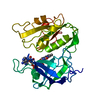 1ndhS S: Starting model for refinement C: citing same article ( |
|---|---|
| Similar structure data |


-Links
 PDBj
PDBj


- Assembly
| Deposited unit | 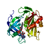
| ||||||||
|---|---|---|---|---|---|---|---|---|---|
| 1 |
| ||||||||
| Unit cell |
|
-Components
| #1: Protein | Mass: 31315.178 Da / Num. of mol.: 1 / Fragment: SOLUBLE DOMAIN Source method: isolated from a genetically manipulated source Source: (gene. exp.)  |
|---|---|
| #2: Chemical | ChemComp-FAD /   Mass: 785.550 Da / Num. of mol.: 1 / Source method: obtained synthetically / Formula: C27H33N9O15P2 / Comment: FAD*YM Mass: 785.550 Da / Num. of mol.: 1 / Source method: obtained synthetically / Formula: C27H33N9O15P2 / Comment: FAD*YM |
| #3: Water | ChemComp-HOH /  Mass: 18.015 Da / Num. of mol.: 148 / Source method: isolated from a natural source / Formula: H2O Mass: 18.015 Da / Num. of mol.: 148 / Source method: isolated from a natural source / Formula: H2O |
-Experimental details
-Experiment
| Experiment | Method: X-RAY DIFFRACTION / Number of used crystals: 1 |
|---|
- Sample preparation
| Crystal | Density Matthews: 3.03 Å3/Da / Density % sol: 59.43 % | ||||||||||||||||||||||||
|---|---|---|---|---|---|---|---|---|---|---|---|---|---|---|---|---|---|---|---|---|---|---|---|---|---|
| Crystal grow | Temperature: 293 K / Method: vapor diffusion, sitting drop / pH: 7.5 Details: PEG 6000, MPD, SODIUM HEPES, pH 7.5, VAPOR DIFFUSION, SITTING DROP, temperature 293K | ||||||||||||||||||||||||
| Crystal grow | *PLUS | ||||||||||||||||||||||||
| Components of the solutions | *PLUS
|
-Data collection
| Diffraction | Mean temperature: 99 K |
|---|---|
| Diffraction source | Source: SYNCHROTRON / Site: NSLS  / Beamline: X12C / Wavelength: 1 Å / Beamline: X12C / Wavelength: 1 Å |
| Detector | Type: BRANDEIS - B4 / Detector: CCD / Date: Oct 11, 2000 / Details: MONOCHROMATOR |
| Radiation | Protocol: SINGLE WAVELENGTH / Monochromatic (M) / Laue (L): M / Scattering type: x-ray |
| Radiation wavelength | Wavelength: 1 Å / Relative weight: 1 |
| Reflection | Resolution: 2→40 Å / Num. all: 26333 / Num. obs: 26290 / % possible obs: 99.8 % / Observed criterion σ(F): 3.2 / Observed criterion σ(I): 11.5 / Redundancy: 6.5 % / Biso Wilson estimate: 13.4 Å2 / Rmerge(I) obs: 0.086 / Net I/σ(I): 11.5 |
| Reflection shell | Resolution: 2→2.07 Å / Redundancy: 5 % / Rmerge(I) obs: 0.216 / Num. unique all: 2530 / % possible all: 99.1 |
| Reflection | *PLUS Redundancy: 6.2 % |
| Reflection shell | *PLUS % possible obs: 99.1 % / Redundancy: 5.3 % / Mean I/σ(I) obs: 3.5 |
- Processing
| Software |
| ||||||||||||||||||||
|---|---|---|---|---|---|---|---|---|---|---|---|---|---|---|---|---|---|---|---|---|---|
| Refinement | Method to determine structure: MOLECULAR REPLACEMENT Starting model: PROTEIN ATOMS OF 1NDH Resolution: 2→30 Å / Cross valid method: THROUGHOUT / σ(F): 0 / σ(I): 0 / Stereochemistry target values: ENGH & HUBER
| ||||||||||||||||||||
| Refinement step | Cycle: LAST / Resolution: 2→30 Å
| ||||||||||||||||||||
| Refine LS restraints |
| ||||||||||||||||||||
| Software | *PLUS Name: CNS / Version: 1 / Classification: refinement | ||||||||||||||||||||
| Refinement | *PLUS Highest resolution: 2 Å / Lowest resolution: 30 Å / σ(F): 0 / Rfactor obs: 0.215 | ||||||||||||||||||||
| Solvent computation | *PLUS | ||||||||||||||||||||
| Displacement parameters | *PLUS | ||||||||||||||||||||
| Refine LS restraints | *PLUS
|
