 Movie
Movie Controller


+ Open data

- Basic information
| Entry | Database: PDB / ID: 1i6t | ||||||
|---|---|---|---|---|---|---|---|
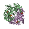
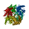
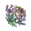
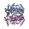
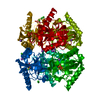
| Title | STRUCTURE OF INORGANIC PYROPHOSPHATASE | ||||||
 Components Components | INORGANIC PYROPHOSPHATASE | ||||||
 Keywords Keywords | HYDROLASE / INORGANIC PYROPHOSPHATASE | ||||||
| Function / homology |  Function and homology information Function and homology informationinorganic triphosphate phosphatase activity / inorganic diphosphatase / inorganic diphosphate phosphatase activity / phosphate-containing compound metabolic process / magnesium ion binding / zinc ion binding / membrane / cytosol Similarity search - Function | ||||||
| Biological species |  | ||||||
| Method |  X-RAY DIFFRACTION / SYNCHROTRON / MOLECULAR REPLACEMENT / Resolution: 1.2 Å X-RAY DIFFRACTION / SYNCHROTRON / MOLECULAR REPLACEMENT / Resolution: 1.2 Å | ||||||
 Authors Authors | Samygina, V.R. / Popov, A.N. / Lamzin, V.S. / Avaeva, S.M. | ||||||
 Citation Citation | Journal: J.Mol.Biol. / Year: 2001 Title: The structures of Escherichia coli inorganic pyrophosphatase complexed with Ca(2+) or CaPP(i) at atomic resolution and their mechanistic implications. Authors: Samygina, V.R. / Popov, A.N. / Rodina, E.V. / Vorobyeva, N.N. / Lamzin, V.S. / Polyakov, K.M. / Kurilova, S.A. / Nazarova, T.I. / Avaeva, S.M. | ||||||
| History |
|
- Structure visualization
| Structure viewer | Molecule: MolmilJmol/JSmol |
|---|
- Downloads & links
-Download
| PDBx/mmCIF format | 1i6t.cif.gz | 99.1 KB | Display | PDBx/mmCIF format |
|---|---|---|---|---|
| PDB format | pdb1i6t.ent.gz | 75.4 KB | Display | PDB format |
| PDBx/mmJSON format | 1i6t.json.gz | Tree view | PDBx/mmJSON format | |
| Others |  Other downloads Other downloads |
-Validation report
| Arichive directory | https://data.pdbj.org/pub/pdb/validation_reports/i6/1i6tftp://data.pdbj.org/pub/pdb/validation_reports/i6/1i6t | HTTPS FTP |
|---|
-Related structure data
| Related structure data |  1i40C  1igpS S: Starting model for refinement C: citing same article ( |
|---|---|
| Similar structure data |


-Links
 PDBj
PDBj


- Assembly
| Deposited unit | 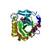
| ||||||||||||
|---|---|---|---|---|---|---|---|---|---|---|---|---|---|
| 1 | x 6
| ||||||||||||
| Unit cell |
| ||||||||||||
| Components on special symmetry positions |
| ||||||||||||
| Details | The biological assembly is a hexamer generated from the monomer in the asymmetric unit by the operation:-y, x-y, z and -x+y, -x, z and y, x, -z and x-y, -y, -z and -x, -x+y, -z |
-Components
-Protein , 1 types, 1 molecules A
| #1: Protein | Mass: 19585.279 Da / Num. of mol.: 1 Source method: isolated from a genetically manipulated source Source: (gene. exp.) |
|---|
-Non-polymers , 5 types, 263 molecules 


| #2: Chemical | ChemComp-CA /  Mass: 40.078 Da / Num. of mol.: 4 / Source method: obtained synthetically / Formula: Ca Mass: 40.078 Da / Num. of mol.: 4 / Source method: obtained synthetically / Formula: Ca#3: Chemical | ChemComp-NA / |  Mass: 22.990 Da / Num. of mol.: 1 / Source method: obtained synthetically / Formula: Na Mass: 22.990 Da / Num. of mol.: 1 / Source method: obtained synthetically / Formula: Na#4: Chemical |  Mass: 35.453 Da / Num. of mol.: 3 / Source method: obtained synthetically / Formula: Cl Mass: 35.453 Da / Num. of mol.: 3 / Source method: obtained synthetically / Formula: Cl#5: Chemical | ChemComp-POP / |  Mass: 175.959 Da / Num. of mol.: 1 / Source method: obtained synthetically / Formula: H2O7P2 Mass: 175.959 Da / Num. of mol.: 1 / Source method: obtained synthetically / Formula: H2O7P2#6: Water | ChemComp-HOH / | Mass: 18.015 Da / Num. of mol.: 254 / Source method: isolated from a natural source / Formula: H2O |
|---|
-Experimental details
-Experiment
| Experiment | Method: X-RAY DIFFRACTION / Number of used crystals: 1 |
|---|
- Sample preparation
| Crystal | Density Matthews: 2.22 Å3/Da / Density % sol: 44.57 % | ||||||||||||||||||||||||||||||||||||||||||||||||||||||||
|---|---|---|---|---|---|---|---|---|---|---|---|---|---|---|---|---|---|---|---|---|---|---|---|---|---|---|---|---|---|---|---|---|---|---|---|---|---|---|---|---|---|---|---|---|---|---|---|---|---|---|---|---|---|---|---|---|---|
| Crystal grow | Temperature: 298 K / Method: vapor diffusion, hanging drop / pH: 5 Details: NULL, pH 5.0, VAPOR DIFFUSION, HANGING DROP, temperature 298.0K | ||||||||||||||||||||||||||||||||||||||||||||||||||||||||
| Crystal grow | *PLUS | ||||||||||||||||||||||||||||||||||||||||||||||||||||||||
| Components of the solutions | *PLUS
|
-Data collection
| Diffraction | Mean temperature: 100 K |
|---|---|
| Diffraction source | Source: SYNCHROTRON / Site: EMBL/DESY, HAMBURG  / Beamline: X11 / Wavelength: 0.906 / Wavelength: 0.906 Å / Beamline: X11 / Wavelength: 0.906 / Wavelength: 0.906 Å |
| Detector | Type: MAR scanner 345 mm plate / Detector: IMAGE PLATE / Date: Nov 22, 1998 / Details: MIRRORS |
| Radiation | Monochromator: SI(111) / Protocol: SINGLE WAVELENGTH / Monochromatic (M) / Laue (L): M / Scattering type: x-ray |
| Radiation wavelength | Wavelength: 0.906 Å / Relative weight: 1 |
| Reflection | Resolution: 1.2→20 Å / Num. all: 640235 / Num. obs: 640235 / % possible obs: 96.1 % / Observed criterion σ(F): 0 / Observed criterion σ(I): 0 / Redundancy: 12.3 % / Biso Wilson estimate: 17.6 Å2 / Rmerge(I) obs: 0.036 / Rsym value: 0.026 / Net I/σ(I): 17.1 |
| Reflection shell | Resolution: 1.2→1.21 Å / Redundancy: 4.8 % / Rmerge(I) obs: 0.441 / Mean I/σ(I) obs: 3.6 / Num. unique all: 1618 / Rsym value: 53.5 / % possible all: 90.1 |
| Reflection | *PLUS Num. obs: 51915 / Num. measured all: 640235 |
| Reflection shell | *PLUS % possible obs: 91.7 % / Rmerge(I) obs: 0.47 |
- Processing
| Software |
| |||||||||||||||||||||||||||||||||
|---|---|---|---|---|---|---|---|---|---|---|---|---|---|---|---|---|---|---|---|---|---|---|---|---|---|---|---|---|---|---|---|---|---|---|
| Refinement | Method to determine structure: MOLECULAR REPLACEMENT Starting model: PDB ENTRY 1IGP Resolution: 1.2→12 Å / Num. parameters: 1487 / Num. restraintsaints: 1830 / σ(F): 0 / σ(I): 0 / Stereochemistry target values: ENGH AND HUBER
| |||||||||||||||||||||||||||||||||
| Refine analyze | Num. disordered residues: 2 / Occupancy sum hydrogen: 1247 / Occupancy sum non hydrogen: 1577 | |||||||||||||||||||||||||||||||||
| Refinement step | Cycle: LAST / Resolution: 1.2→12 Å
| |||||||||||||||||||||||||||||||||
| Refine LS restraints |
| |||||||||||||||||||||||||||||||||
| Software | *PLUS Name: SHELXL-97 / Classification: refinement | |||||||||||||||||||||||||||||||||
| Refinement | *PLUS σ(F): 0 / Rfactor Rfree: 0.17 | |||||||||||||||||||||||||||||||||
| Solvent computation | *PLUS | |||||||||||||||||||||||||||||||||
| Displacement parameters | *PLUS | |||||||||||||||||||||||||||||||||
| Refine LS restraints | *PLUS
|
