 Movie
Movie Controller


+ Open data

- Basic information
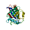
| Entry | Database: PDB / ID: 1i5g | ||||||
|---|---|---|---|---|---|---|---|
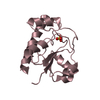
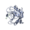
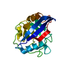
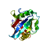
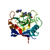
| Title | TRYPAREDOXIN II COMPLEXED WITH GLUTATHIONYLSPERMIDINE | ||||||
 Components Components | TRYPAREDOXIN II | ||||||
 Keywords Keywords | ELECTRON TRANSPORT / tryparedoxin | ||||||
| Function / homology |  Function and homology information Function and homology informationthioredoxin-disulfide reductase (NADPH) activity / negative regulation of Wnt signaling pathway / negative regulation of protein ubiquitination / nucleus Similarity search - Function | ||||||
| Biological species |  Crithidia fasciculata (eukaryote) Crithidia fasciculata (eukaryote) | ||||||
| Method |  X-RAY DIFFRACTION / SYNCHROTRON / MOLECULAR REPLACEMENT / Resolution: 1.4 Å X-RAY DIFFRACTION / SYNCHROTRON / MOLECULAR REPLACEMENT / Resolution: 1.4 Å | ||||||
 Authors Authors | Hofmann, B. / Budde, H. / Bruns, K. / Guerrero, S.A. / Kalisz, H.M. / Menge, U. / Montemartini, M. / Nogoceke, E. / Steinert, P. / Wissing, J.B. ...Hofmann, B. / Budde, H. / Bruns, K. / Guerrero, S.A. / Kalisz, H.M. / Menge, U. / Montemartini, M. / Nogoceke, E. / Steinert, P. / Wissing, J.B. / Flohe, L. / Hecht, H.-J. | ||||||
 Citation Citation | Journal: Biol.Chem. / Year: 2001 Title: Structures of tryparedoxins revealing interaction with trypanothione. Authors: Hofmann, B. / Budde, H. / Bruns, K. / Guerrero, S.A. / Kalisz, H.M. / Menge, U. / Montemartini, M. / Nogoceke, E. / Steinert, P. / Wissing, J.B. / Flohe, L. / Hecht, H.J. | ||||||
| History |
|
- Structure visualization
| Structure viewer | Molecule: MolmilJmol/JSmol |
|---|
- Downloads & links
-Download
| PDBx/mmCIF format | 1i5g.cif.gz | 50.5 KB | Display | PDBx/mmCIF format |
|---|---|---|---|---|
| PDB format | pdb1i5g.ent.gz | 34.4 KB | Display | PDB format |
| PDBx/mmJSON format | 1i5g.json.gz | Tree view | PDBx/mmJSON format | |
| Others |  Other downloads Other downloads |
-Validation report
| Arichive directory | https://data.pdbj.org/pub/pdb/validation_reports/i5/1i5gftp://data.pdbj.org/pub/pdb/validation_reports/i5/1i5g | HTTPS FTP |
|---|
-Related structure data
| Related structure data | 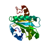 1ewxC 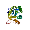 1ezkC 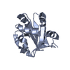 1fg4SC S: Starting model for refinement C: citing same article ( |
|---|---|
| Similar structure data |


-Links
 PDBj
PDBj- Assembly
| Deposited unit | 
| ||||||||
|---|---|---|---|---|---|---|---|---|---|
| 1 |
| ||||||||
| Unit cell |
|
-Components
| #1: Protein | Mass: 16223.512 Da / Num. of mol.: 1 / Mutation: C44S Source method: isolated from a genetically manipulated source Source: (gene. exp.) Crithidia fasciculata (eukaryote) / Strain: HS6 / Plasmid: PET22B / Production host:  |
|---|---|
| #2: Chemical | ChemComp-TRS /   Mass: 122.143 Da / Num. of mol.: 1 / Source method: obtained synthetically / Formula: C4H12NO3 / Comment: pH buffer*YM Mass: 122.143 Da / Num. of mol.: 1 / Source method: obtained synthetically / Formula: C4H12NO3 / Comment: pH buffer*YM |
| #3: Chemical | ChemComp-TS5 /   Mass: 434.554 Da / Num. of mol.: 1 / Source method: obtained synthetically / Formula: C17H34N6O5S Mass: 434.554 Da / Num. of mol.: 1 / Source method: obtained synthetically / Formula: C17H34N6O5S |
| #4: Water | ChemComp-HOH /  Mass: 18.015 Da / Num. of mol.: 279 / Source method: isolated from a natural source / Formula: H2O Mass: 18.015 Da / Num. of mol.: 279 / Source method: isolated from a natural source / Formula: H2O |
| Has protein modification | Y |
-Experimental details
-Experiment
| Experiment | Method: X-RAY DIFFRACTION / Number of used crystals: 1 |
|---|
- Sample preparation
| Crystal | Density Matthews: 2.16 Å3/Da / Density % sol: 42.95 % | ||||||||||||||||||||||||||||||
|---|---|---|---|---|---|---|---|---|---|---|---|---|---|---|---|---|---|---|---|---|---|---|---|---|---|---|---|---|---|---|---|
| Crystal grow | Temperature: 292 K / Method: vapor diffusion, sitting drop / pH: 6.9 Details: PEG 8000, Mes Tris glutathionylspermidine, pH 6.9, VAPOR DIFFUSION, SITTING DROP, temperature 292K | ||||||||||||||||||||||||||||||
| Crystal grow | *PLUS Temperature: 19 ℃ | ||||||||||||||||||||||||||||||
| Components of the solutions | *PLUS
|
-Data collection
| Diffraction | Mean temperature: 100 K |
|---|---|
| Diffraction source | Source: SYNCHROTRON / Site: MPG/DESY, HAMBURG  / Beamline: BW6 / Wavelength: 0.95 Å / Beamline: BW6 / Wavelength: 0.95 Å |
| Detector | Type: MARRESEARCH / Detector: CCD / Date: Sep 25, 2000 / Details: mirrors |
| Radiation | Monochromator: Si 111 / Protocol: SINGLE WAVELENGTH / Monochromatic (M) / Laue (L): M / Scattering type: x-ray |
| Radiation wavelength | Wavelength: 0.95 Å / Relative weight: 1 |
| Reflection | Resolution: 1.4→28.87 Å / Num. all: 24911 / Num. obs: 24911 / % possible obs: 89 % / Observed criterion σ(F): 0 / Observed criterion σ(I): 0 / Redundancy: 4.3 % / Biso Wilson estimate: 9.2 Å2 / Rmerge(I) obs: 0.074 / Rsym value: 0.065 / Net I/σ(I): 4.6 |
| Reflection shell | Resolution: 1.4→1.48 Å / Redundancy: 4.1 % / Rmerge(I) obs: 0.088 / Mean I/σ(I) obs: 8.8 / Num. unique all: 3735 / Rsym value: 0.077 / % possible all: 92.1 |
| Reflection | *PLUS Num. obs: 24680 / % possible obs: 89 % / Rmerge(I) obs: 0.065 |
- Processing
| Software |
| |||||||||||||||||||||||||
|---|---|---|---|---|---|---|---|---|---|---|---|---|---|---|---|---|---|---|---|---|---|---|---|---|---|---|
| Refinement | Method to determine structure: MOLECULAR REPLACEMENT Starting model: PDB ENTRY 1FG4 Resolution: 1.4→40.49 Å / Isotropic thermal model: Isotropic / Cross valid method: THROUGHOUT / σ(F): 0 / σ(I): 0 / Stereochemistry target values: Engh & Huber
| |||||||||||||||||||||||||
| Displacement parameters | Biso mean: 7 Å2
| |||||||||||||||||||||||||
| Refine analyze | Luzzati coordinate error free: 0.075 Å | |||||||||||||||||||||||||
| Refinement step | Cycle: LAST / Resolution: 1.4→40.49 Å
| |||||||||||||||||||||||||
| Refine LS restraints |
| |||||||||||||||||||||||||
| Software | *PLUS Name: REFMAC / Version: 5 / Classification: refinement | |||||||||||||||||||||||||
| Refinement | *PLUS σ(F): 0 / % reflection Rfree: 4.9 % / Rfactor obs: 0.176 | |||||||||||||||||||||||||
| Solvent computation | *PLUS | |||||||||||||||||||||||||
| Displacement parameters | *PLUS |
