 Movie
Movie Controller


+ Open data

- Basic information
| Entry | Database: PDB / ID: 1i48 | ||||||
|---|---|---|---|---|---|---|---|
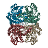
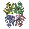
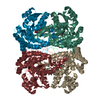
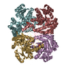
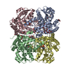
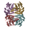
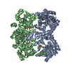
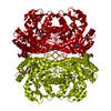
| Title | CYSTATHIONINE GAMMA-SYNTHASE IN COMPLEX WITH THE INHIBITOR CTCPO | ||||||
 Components Components | CYSTATHIONINE GAMMA-SYNTHASE | ||||||
 Keywords Keywords | LYASE / PLP-dependent enzyme / homotetramer / inhibitor complex / CTCPO | ||||||
| Function / homology |  Function and homology information Function and homology informationplant cystathionine gamma-synthase / cystathionine gamma-synthase activity / transsulfuration / methionine biosynthetic process / pyridoxal phosphate binding Similarity search - Function | ||||||
| Biological species |  | ||||||
| Method |  X-RAY DIFFRACTION / MOLECULAR REPLACEMENT / Resolution: 3.25 Å X-RAY DIFFRACTION / MOLECULAR REPLACEMENT / Resolution: 3.25 Å | ||||||
 Authors Authors | Steegborn, C. / Laber, B. / Messerschmidt, A. / Huber, R. / Clausen, T. | ||||||
 Citation Citation | Journal: J.Mol.Biol. / Year: 2001 Title: Crystal structures of cystathionine gamma-synthase inhibitor complexes rationalize the increased affinity of a novel inhibitor. Authors: Steegborn, C. / Laber, B. / Messerschmidt, A. / Huber, R. / Clausen, T. | ||||||
| History |
|
- Structure visualization
| Structure viewer | Molecule: MolmilJmol/JSmol |
|---|
- Downloads & links
-Download
| PDBx/mmCIF format | 1i48.cif.gz | 882.5 KB | Display | PDBx/mmCIF format |
|---|---|---|---|---|
| PDB format | pdb1i48.ent.gz | 738.4 KB | Display | PDB format |
| PDBx/mmJSON format | 1i48.json.gz | Tree view | PDBx/mmJSON format | |
| Others |  Other downloads Other downloads |
-Validation report
| Summary document | 1i48_validation.pdf.gz | 931.3 KB | Display | wwPDB validaton report |
|---|---|---|---|---|
| Full document | 1i48_full_validation.pdf.gz | 1.1 MB | Display | |
| Data in XML | 1i48_validation.xml.gz | 116.1 KB | Display | |
| Data in CIF | 1i48_validation.cif.gz | 163.3 KB | Display | |
| Arichive directory | https://data.pdbj.org/pub/pdb/validation_reports/i4/1i48ftp://data.pdbj.org/pub/pdb/validation_reports/i4/1i48 | HTTPS FTP |
-Related structure data
| Related structure data |  1i41C  1i43C  1qgnS S: Starting model for refinement C: citing same article ( |
|---|---|
| Similar structure data |


-Links
 PDBj
PDBj- Assembly
| Deposited unit | 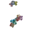
| ||||||||
|---|---|---|---|---|---|---|---|---|---|
| 1 | 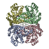
| ||||||||
| 2 | 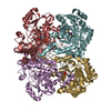
| ||||||||
| 3 | 
| ||||||||
| Unit cell |
|
-Components
| #1: Protein | Mass: 48067.973 Da / Num. of mol.: 12 Source method: isolated from a genetically manipulated source Source: (gene. exp.)  #2: Chemical | ChemComp-PLP /   Mass: 247.142 Da / Num. of mol.: 12 / Source method: obtained synthetically / Formula: C8H10NO6P Mass: 247.142 Da / Num. of mol.: 12 / Source method: obtained synthetically / Formula: C8H10NO6P#3: Chemical | ChemComp-CCO /   Mass: 270.692 Da / Num. of mol.: 12 / Source method: obtained synthetically / Formula: C10H7ClN2O3S Mass: 270.692 Da / Num. of mol.: 12 / Source method: obtained synthetically / Formula: C10H7ClN2O3S |
|---|
-Experimental details
-Experiment
| Experiment | Method: X-RAY DIFFRACTION / Number of used crystals: 1 |
|---|
- Sample preparation
| Crystal | Density Matthews: 3.64 Å3/Da / Density % sol: 66.17 % | ||||||||||||||||||||||||||||||
|---|---|---|---|---|---|---|---|---|---|---|---|---|---|---|---|---|---|---|---|---|---|---|---|---|---|---|---|---|---|---|---|
| Crystal grow | Temperature: 291 K / Method: vapor diffusion / pH: 6.5 Details: MES, magnesium acetate, PEG 8000, pH 6.5, VAPOR DIFFUSION, temperature 291K | ||||||||||||||||||||||||||||||
| Crystal grow | *PLUS pH: 7.8 / Method: vapor diffusion, sitting drop / Details: used seeding | ||||||||||||||||||||||||||||||
| Components of the solutions | *PLUS
|
-Data collection
| Diffraction | Mean temperature: 291 K |
|---|---|
| Diffraction source | Source: ROTATING ANODE / Type: RIGAKU RU200 / Wavelength: 1.5418 Å |
| Detector | Type: MARRESEARCH / Detector: IMAGE PLATE / Date: Jul 10, 1999 |
| Radiation | Protocol: SINGLE WAVELENGTH / Monochromatic (M) / Laue (L): M / Scattering type: x-ray |
| Radiation wavelength | Wavelength: 1.5418 Å / Relative weight: 1 |
| Reflection | Resolution: 3.25→20 Å / Num. all: 119179 / Num. obs: 119179 / % possible obs: 93 % / Observed criterion σ(I): -100 / Redundancy: 1.7 % / Rmerge(I) obs: 0.138 / Net I/σ(I): 4.4 |
| Reflection shell | Resolution: 3.25→3.36 Å / Redundancy: 1.6 % / Rmerge(I) obs: 0.496 / % possible all: 94 |
| Reflection | *PLUS % possible obs: 93 % |
| Reflection shell | *PLUS % possible obs: 94 % |
- Processing
| Software |
| ||||||||||||||||||||
|---|---|---|---|---|---|---|---|---|---|---|---|---|---|---|---|---|---|---|---|---|---|
| Refinement | Method to determine structure: MOLECULAR REPLACEMENT Starting model: PDB entry 1qgn Resolution: 3.25→20 Å / Isotropic thermal model: Isotropic / Cross valid method: THROUGHOUT / σ(F): 2 / Stereochemistry target values: Engh & Huber
| ||||||||||||||||||||
| Refinement step | Cycle: LAST / Resolution: 3.25→20 Å
| ||||||||||||||||||||
| Refine LS restraints |
| ||||||||||||||||||||
| Software | *PLUS Name: X-PLOR / Classification: refinement | ||||||||||||||||||||
| Refinement | *PLUS Lowest resolution: 20 Å / σ(F): 2 / Rfactor obs: 0.226 | ||||||||||||||||||||
| Solvent computation | *PLUS | ||||||||||||||||||||
| Displacement parameters | *PLUS | ||||||||||||||||||||
| Refine LS restraints | *PLUS
|
