 Movie
Movie Controller

[English] 日本語
 Yorodumi
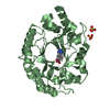
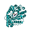
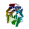
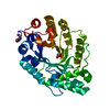
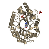
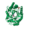
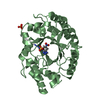
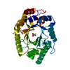
- PDB-1i1x: 1.11 A ATOMIC RESOLUTION STRUCTURE OF A THERMOSTABLE XYLANASE FRO... -
+ Open data

- Basic information
| Entry | Database: PDB / ID: 1i1x | |||||||||
|---|---|---|---|---|---|---|---|---|---|---|
| Title | 1.11 A ATOMIC RESOLUTION STRUCTURE OF A THERMOSTABLE XYLANASE FROM THERMOASCUS AURANTIACUS | |||||||||
 Components Components | ENDO-1,4-BETA-XYLANASE | |||||||||
 Keywords Keywords | HYDROLASE / XYLAN DEGRADATION / GLYCOSIDASE / ENZYME / ATOMIC RESOLUTION / ROOM TEMPERATURE / 1 / 4-BETA-XYLAN XYLANOHYDROLASE | |||||||||
| Function / homology |  Function and homology information Function and homology informationendo-1,4-beta-xylanase / endo-1,4-beta-xylanase activity / xylan catabolic process Similarity search - Function | |||||||||
| Biological species |  Thermoascus aurantiacus (fungus) Thermoascus aurantiacus (fungus) | |||||||||
| Method |  X-RAY DIFFRACTION / SYNCHROTRON / MOLECULAR REPLACEMENT / Resolution: 1.11 Å X-RAY DIFFRACTION / SYNCHROTRON / MOLECULAR REPLACEMENT / Resolution: 1.11 Å | |||||||||
 Authors Authors | Natesh, R. / Ramakumar, S. / Viswamitra, M.A. | |||||||||
 Citation Citation | Journal: Acta Crystallogr.,Sect.D / Year: 2003 Title: Thermostable xylanase from Thermoascus aurantiacus at ultrahigh resolution (0.89 A) at 100 K and atomic resolution (1.11 A) at 293 K refined anisotropically to small-molecule accuracy. Authors: Natesh, R. / Manikandan, K. / Bhanumoorthy, P. / Viswamitra, M.A. / Ramakumar, S. #1: Journal: J.Mol.Biol. / Year: 1999Title: Crystal Structure at 1.8 A Resolution and Proposed Amino Acid Sequence of a Thermostable Xylanase from Thermoascus Aurantiascus Authors: Natesh, R. / Bhanumoorthy, P. / Vithayathil, P.J. / Sekar, K. / Ramakumar, S. / Viswamitra, M.A. #2: Journal: J.Mol.Biol. / Year: 1993Title: Crystallization and Preliminary X-Ray Diffraction Analysis of Crystals of Thermoascus Aurantiacus Xylanase Authors: Viswamitra, M.A. / Bhanumoorthy, P. / Ramakumar, S. / Manjula, M.V. / Vithayathil, P.J. / Murthy, S.K. / Naren, A.P. | |||||||||
| History |
|
- Structure visualization
| Structure viewer | Molecule: MolmilJmol/JSmol |
|---|
- Downloads & links
-Download
| PDBx/mmCIF format | 1i1x.cif.gz | 181.6 KB | Display | PDBx/mmCIF format |
|---|---|---|---|---|
| PDB format | pdb1i1x.ent.gz | 146.2 KB | Display | PDB format |
| PDBx/mmJSON format | 1i1x.json.gz | Tree view | PDBx/mmJSON format | |
| Others |  Other downloads Other downloads |
-Validation report
| Arichive directory | https://data.pdbj.org/pub/pdb/validation_reports/i1/1i1xftp://data.pdbj.org/pub/pdb/validation_reports/i1/1i1x | HTTPS FTP |
|---|
-Related structure data
| Related structure data | 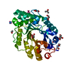 1i1wC 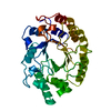 1tuxS S: Starting model for refinement C: citing same article ( |
|---|---|
| Similar structure data |


-Links
 PDBj
PDBj
- Assembly
| Deposited unit | 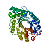
| ||||||||
|---|---|---|---|---|---|---|---|---|---|
| 1 |
| ||||||||
| Unit cell |
|
-Components
| #1: Protein | Mass: 32868.734 Da / Num. of mol.: 1 / Source method: isolated from a natural source / Source: (natural) Thermoascus aurantiacus (fungus) / Strain: STRAIN ISOLATED FROM LOCAL INDIAN SOIL / References: UniProt: P23360, endo-1,4-beta-xylanase |
|---|---|
| #2: Water | ChemComp-HOH /  Mass: 18.015 Da / Num. of mol.: 265 / Source method: isolated from a natural source / Formula: H2O Mass: 18.015 Da / Num. of mol.: 265 / Source method: isolated from a natural source / Formula: H2O |
| Has protein modification | Y |
-Experimental details
-Experiment
| Experiment | Method: X-RAY DIFFRACTION / Number of used crystals: 1 |
|---|
- Sample preparation
| Crystal | Density Matthews: 2.03 Å3/Da / Density % sol: 39.32 % | ||||||||||||||||||||
|---|---|---|---|---|---|---|---|---|---|---|---|---|---|---|---|---|---|---|---|---|---|
| Crystal grow | Temperature: 293 K / Method: vapor diffusion, sitting drop / pH: 7.2 Details: CRYSTALS WERE GROWN BY SITTING DROP AGAINST 12% AMMONIUM SULPHATE SOLUTION AT THE SAME BUFFER CONDITION IN THE RESERVOIR., pH 7.2, VAPOR DIFFUSION, SITTING DROP, temperature 293K | ||||||||||||||||||||
| Crystal grow | *PLUS Method: vapor diffusion, hanging drop / Details: Viswamitra, M.A., (1993) J.Mol.Biol., 232, 987. | ||||||||||||||||||||
| Components of the solutions | *PLUS
|
-Data collection
| Diffraction | Mean temperature: 293 K |
|---|---|
| Diffraction source | Source: SYNCHROTRON / Site: NSLS  / Beamline: X9B / Wavelength: 1.009 / Wavelength: 1.009 Å / Beamline: X9B / Wavelength: 1.009 / Wavelength: 1.009 Å |
| Detector | Type: ADSC QUANTUM 4 / Detector: CCD / Date: Feb 28, 1999 / Details: MIRROR |
| Radiation | Monochromator: DOUBLE CRYSTAL MONOCHROMATOR / Protocol: SINGLE WAVELENGTH / Monochromatic (M) / Laue (L): M / Scattering type: x-ray |
| Radiation wavelength | Wavelength: 1.009 Å / Relative weight: 1 |
| Reflection | Resolution: 1.11→25 Å / Num. obs: 90855 / % possible obs: 87.5 % / Redundancy: 3.2 % / Rmerge(I) obs: 0.052 / Net I/σ(I): 20.6 |
| Reflection shell | Resolution: 1.11→1.15 Å / Rmerge(I) obs: 0.36 / Mean I/σ(I) obs: 3 / % possible all: 81.3 |
| Reflection | *PLUS Redundancy: 3.25 % / Num. measured all: 294946 |
| Reflection shell | *PLUS % possible obs: 81.3 % / Rmerge(I) obs: 0.36 / Mean I/σ(I) obs: 2.98 |
- Processing
| Software |
| |||||||||||||||||||||||||||||||||
|---|---|---|---|---|---|---|---|---|---|---|---|---|---|---|---|---|---|---|---|---|---|---|---|---|---|---|---|---|---|---|---|---|---|---|
| Refinement | Method to determine structure: MOLECULAR REPLACEMENT Starting model: 1TUX (1.8 A) Resolution: 1.11→10 Å / Num. parameters: 22784 / Num. restraintsaints: 28345 / Cross valid method: FREE R / σ(F): 0 / Stereochemistry target values: ENGH AND HUBER / Details: CNS 0.4 was also used for refinement
| |||||||||||||||||||||||||||||||||
| Solvent computation | Solvent model: MOEWS & KRETSINGER, J.MOL.BIOL.91(1973)201-228 | |||||||||||||||||||||||||||||||||
| Refine analyze | Num. disordered residues: 29 / Occupancy sum hydrogen: 2119.2 / Occupancy sum non hydrogen: 2536.06 | |||||||||||||||||||||||||||||||||
| Refinement step | Cycle: LAST / Resolution: 1.11→10 Å
| |||||||||||||||||||||||||||||||||
| Refine LS restraints |
| |||||||||||||||||||||||||||||||||
| Software | *PLUS Name: SHELXL / Version: 97 / Classification: refinement | |||||||||||||||||||||||||||||||||
| Refinement | *PLUS Lowest resolution: 10 Å | |||||||||||||||||||||||||||||||||
| Solvent computation | *PLUS | |||||||||||||||||||||||||||||||||
| Displacement parameters | *PLUS | |||||||||||||||||||||||||||||||||
| Refine LS restraints | *PLUS
|
