 Movie
Movie Controller

[English] 日本語
 Yorodumi
- PDB-1hzk: SOLUTION STRUCTURES OF C-1027 APOPROTEIN AND ITS COMPLEX WITH THE... -
+ Open data

- Basic information
| Entry | Database: PDB / ID: 1hzk | ||||||
|---|---|---|---|---|---|---|---|
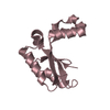
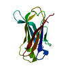
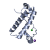
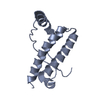
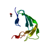
| Title | SOLUTION STRUCTURES OF C-1027 APOPROTEIN AND ITS COMPLEX WITH THE AROMATIZED CHROMOPHORE | ||||||
 Components Components | C-1027 APOPROTEIN | ||||||
 Keywords Keywords | ANTIBIOTIC / chromoprotein / C-1027 / apoprotein | ||||||
| Function / homology |  Function and homology information Function and homology information | ||||||
| Biological species |  Streptomyces globisporus (bacteria) Streptomyces globisporus (bacteria) | ||||||
| Method | SOLUTION NMR / simulated annealing | ||||||
 Authors Authors | Tanaka, T. / Fukuda-Ishisaka, S. / Hirama, M. / Otani, T. | ||||||
 Citation Citation | Journal: J.Mol.Biol. / Year: 2001 Title: Solution structures of C-1027 apoprotein and its complex with the aromatized chromophore. Authors: Tanaka, T. / Fukuda-Ishisaka, S. / Hirama, M. / Otani, T. | ||||||
| History |
|
- Structure visualization
| Structure viewer | Molecule: MolmilJmol/JSmol |
|---|
- Downloads & links
-Download
| PDBx/mmCIF format | 1hzk.cif.gz | 819.9 KB | Display | PDBx/mmCIF format |
|---|---|---|---|---|
| PDB format | pdb1hzk.ent.gz | 685.5 KB | Display | PDB format |
| PDBx/mmJSON format | 1hzk.json.gz | Tree view | PDBx/mmJSON format | |
| Others |  Other downloads Other downloads |
-Validation report
| Arichive directory | https://data.pdbj.org/pub/pdb/validation_reports/hz/1hzkftp://data.pdbj.org/pub/pdb/validation_reports/hz/1hzk | HTTPS FTP |
|---|
-Related structure data
| Related structure data |  1hzlC C: citing same article ( |
|---|---|
| Similar structure data | |
| Other databases |
|


-Links
 PDBj
PDBj- Assembly
| Deposited unit | 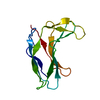
| |||||||||
|---|---|---|---|---|---|---|---|---|---|---|
| 1 |
| |||||||||
| NMR ensembles |
|
-Components
| #1: Protein | Mass: 10504.340 Da / Num. of mol.: 1 / Source method: isolated from a natural source / Source: (natural) Streptomyces globisporus (bacteria) / Strain: C-1027 / References: UniProt: Q06110 |
|---|---|
| Has protein modification | Y |
-Experimental details
-Experiment
| Experiment | Method: SOLUTION NMR | ||||||||||||||||||||
|---|---|---|---|---|---|---|---|---|---|---|---|---|---|---|---|---|---|---|---|---|---|
| NMR experiment |
| ||||||||||||||||||||
| NMR details | Text: This structure was determined using standard 2D homonuclear techniques. |
- Sample preparation
| Details | Contents: Sample 1: 6.6-7.6mM C-1027 apoprotein; 99.996% D2O; Sample 2: 6.6-7.6mM C-1027 apoprotein; 90% H2O, 10% D2O |
|---|---|
| Sample conditions | pH: 5 / Pressure: ambient / Temperature: 303 K |
| Crystal grow | *PLUS Method: other / Details: NMR |
-NMR measurement
| NMR spectrometer | Type: Bruker AM / Manufacturer: Bruker / Model: AM / Field strength: 600 MHz |
|---|
- Processing
| NMR software |
| ||||||||||||
|---|---|---|---|---|---|---|---|---|---|---|---|---|---|
| Refinement | Method: simulated annealing / Software ordinal: 1 Details: The structures are based on a total of 1539 restraints: 1383 NOE-derived distance restraints, 86 dihedral angle restraints, 64 distance restraints from hydrogen bonds, and 6 distance ...Details: The structures are based on a total of 1539 restraints: 1383 NOE-derived distance restraints, 86 dihedral angle restraints, 64 distance restraints from hydrogen bonds, and 6 distance restraints from disulfide bonds. | ||||||||||||
| NMR representative | Selection criteria: lowest energy | ||||||||||||
| NMR ensemble | Conformer selection criteria: structures with the lowest energy Conformers calculated total number: 100 / Conformers submitted total number: 30 |
 X-PLOR