 Movie
Movie Controller


+ Open data

- Basic information
| Entry | Database: PDB / ID: 1hwp | |||||||||
|---|---|---|---|---|---|---|---|---|---|---|
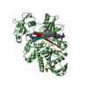
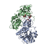
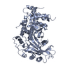
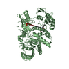
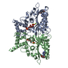
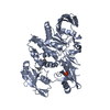
| Title | EBULIN COMPLEXED WITH PTEROIC ACID, TRIGONAL CRYSTAL FORM | |||||||||
 Components Components | (EBULIN) x 2 | |||||||||
 Keywords Keywords | HYDROLASE / Ribosome-inactivating protein / ricin-like / inhibitor | |||||||||
| Function / homology |  Function and homology information Function and homology informationrRNA N-glycosylase / rRNA N-glycosylase activity / defense response / toxin activity / negative regulation of translation Similarity search - Function | |||||||||
| Biological species |  Sambucus ebulus (plant) Sambucus ebulus (plant) | |||||||||
| Method |  X-RAY DIFFRACTION / MOLECULAR REPLACEMENT / Resolution: 3.1 Å X-RAY DIFFRACTION / MOLECULAR REPLACEMENT / Resolution: 3.1 Å | |||||||||
 Authors Authors | Pascal, J.M. / Day, P.J. / Monzingo, A.F. / Ernst, S.R. / Robertus, J.D. | |||||||||
 Citation Citation | Journal: Proteins / Year: 2001 Title: 2.8-A crystal structure of a nontoxic type-II ribosome-inactivating protein, ebulin l. Authors: Pascal, J.M. / Day, P.J. / Monzingo, A.F. / Ernst, S.R. / Robertus, J.D. / Iglesias, R. / Perez, Y. / Ferreras, J.M. / Citores, L. / Girbes, T. | |||||||||
| History |
|
- Structure visualization
| Structure viewer | Molecule: MolmilJmol/JSmol |
|---|
- Downloads & links
-Download
| PDBx/mmCIF format | 1hwp.cif.gz | 107.1 KB | Display | PDBx/mmCIF format |
|---|---|---|---|---|
| PDB format | pdb1hwp.ent.gz | 82.5 KB | Display | PDB format |
| PDBx/mmJSON format | 1hwp.json.gz | Tree view | PDBx/mmJSON format | |
| Others |  Other downloads Other downloads |
-Validation report
| Arichive directory | https://data.pdbj.org/pub/pdb/validation_reports/hw/1hwpftp://data.pdbj.org/pub/pdb/validation_reports/hw/1hwp | HTTPS FTP |
|---|
-Related structure data
| Related structure data |  1hwmC  1hwnC  1hwoSC C: citing same article ( S: Starting model for refinement |
|---|---|
| Similar structure data |


-Links
 PDBj
PDBj


- Assembly
| Deposited unit | 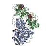
| ||||||||
|---|---|---|---|---|---|---|---|---|---|
| 1 |
| ||||||||
| 2 | 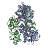
| ||||||||
| Unit cell |
|
-Components
-Protein , 2 types, 2 molecules AB
| #1: Protein | Mass: 28478.982 Da / Num. of mol.: 1 / Source method: isolated from a natural source / Details: N-GLYCOSIDASE / Source: (natural) Sambucus ebulus (plant) / Tissue: LEAF / References: UniProt: Q9AVR2, rRNA N-glycosylase |
|---|---|
| #2: Protein | Mass: 29377.898 Da / Num. of mol.: 1 / Source method: isolated from a natural source / Details: GALACTOSIDE SPECIFIC LECTIN / Source: (natural) Sambucus ebulus (plant) / Tissue: LEAF / References: UniProt: Q9AVR2, rRNA N-glycosylase |
-Sugars , 2 types, 2 molecules
| #3: Polysaccharide | beta-D-galactopyranose-(1-4)-beta-D-glucopyranose / beta-lactose  Source method: isolated from a genetically manipulated source Details: oligosaccharide / References: beta-lactose |
|---|---|
| #4: Polysaccharide | beta-D-mannopyranose-(1-4)-2-acetamido-2-deoxy-beta-D-glucopyranose-(1-4)-2-acetamido-2-deoxy-beta- ...beta-D-mannopyranose-(1-4)-2-acetamido-2-deoxy-beta-D-glucopyranose-(1-4)-2-acetamido-2-deoxy-beta-D-glucopyranose Source method: isolated from a genetically manipulated source |
-Non-polymers , 2 types, 21 molecules 

| #5: Chemical | ChemComp-PT1 /  Mass: 312.283 Da / Num. of mol.: 1 / Source method: obtained synthetically / Formula: C14H12N6O3 Mass: 312.283 Da / Num. of mol.: 1 / Source method: obtained synthetically / Formula: C14H12N6O3 |
|---|---|
| #6: Water | ChemComp-HOH / Mass: 18.015 Da / Num. of mol.: 20 / Source method: isolated from a natural source / Formula: H2O |
-Details
| Has protein modification | Y |
|---|
-Experimental details
-Experiment
| Experiment | Method: X-RAY DIFFRACTION / Number of used crystals: 1 |
|---|
- Sample preparation
| Crystal | Density Matthews: 2.62 Å3/Da / Density % sol: 53.13 % | ||||||||||||||||||||
|---|---|---|---|---|---|---|---|---|---|---|---|---|---|---|---|---|---|---|---|---|---|
| Crystal grow | Temperature: 298 K / Method: vapor diffusion, hanging drop / pH: 7.5 Details: 1.1 M Na,K Tartrate, 100 mM HEPES, pH 7.5, VAPOR DIFFUSION, HANGING DROP, temperature 298K | ||||||||||||||||||||
| Crystal grow | *PLUS Temperature: 25 ℃ | ||||||||||||||||||||
| Components of the solutions | *PLUS
|
-Data collection
| Diffraction | Mean temperature: 150 K |
|---|---|
| Diffraction source | Source: ROTATING ANODE / Type: RIGAKU RU200 / Wavelength: 1.54 Å |
| Detector | Type: RIGAKU RAXIS IV / Detector: IMAGE PLATE / Date: Oct 21, 1998 |
| Radiation | Monochromator: Double focusing mirrors, (Ni + Pt) + Ni filters Protocol: SINGLE WAVELENGTH / Monochromatic (M) / Laue (L): M / Scattering type: x-ray |
| Radiation wavelength | Wavelength: 1.54 Å / Relative weight: 1 |
| Reflection | Resolution: 3.1→40 Å / Num. all: 11311 / Num. obs: 11311 / % possible obs: 97.5 % / Observed criterion σ(F): 0 / Observed criterion σ(I): 0 / Redundancy: 1.95 % / Rmerge(I) obs: 0.118 / Net I/σ(I): 7.3 |
| Reflection shell | Resolution: 3.1→3.21 Å / Rmerge(I) obs: 0.378 / % possible all: 98.1 |
| Reflection shell | *PLUS % possible obs: 98.1 % / Mean I/σ(I) obs: 2.1 |
- Processing
| Software |
| ||||||||||||||||||||
|---|---|---|---|---|---|---|---|---|---|---|---|---|---|---|---|---|---|---|---|---|---|
| Refinement | Method to determine structure: MOLECULAR REPLACEMENT Starting model: PDB entry 1HWO Resolution: 3.1→40 Å / σ(F): 2 / Stereochemistry target values: Engh & Huber
| ||||||||||||||||||||
| Refinement step | Cycle: LAST / Resolution: 3.1→40 Å
| ||||||||||||||||||||
| Refine LS restraints |
| ||||||||||||||||||||
| Software | *PLUS Name: X-PLOR / Version: 3.851 / Classification: refinement | ||||||||||||||||||||
| Refine LS restraints | *PLUS
|
