 Movie
Movie Controller


+ Open data

- Basic information
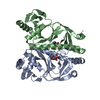
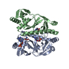
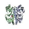
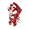
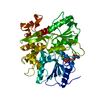
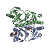
| Entry | Database: PDB / ID: 1hw5 | ||||||
|---|---|---|---|---|---|---|---|
| Title | THE CAP/CRP VARIANT T127L/S128A | ||||||
 Components Components | CATABOLITE GENE ACTIVATOR | ||||||
 Keywords Keywords | GENE REGULATION / cAMP Receptor Protein / Catabolite Activator Protein (CAP) Transcription / Allostery / cAMP / cyclic AMP Mutant | ||||||
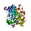
| Function / homology |  Function and homology information Function and homology informationcarbon catabolite repression of transcription / DNA binding, bending / minor groove of adenine-thymine-rich DNA binding / cAMP binding / protein-DNA complex / sequence-specific DNA binding / DNA-binding transcription factor activity / negative regulation of DNA-templated transcription / positive regulation of DNA-templated transcription / DNA-templated transcription ...carbon catabolite repression of transcription / DNA binding, bending / minor groove of adenine-thymine-rich DNA binding / cAMP binding / protein-DNA complex / sequence-specific DNA binding / DNA-binding transcription factor activity / negative regulation of DNA-templated transcription / positive regulation of DNA-templated transcription / DNA-templated transcription / identical protein binding / cytosol Similarity search - Function | ||||||
| Biological species |  | ||||||
| Method |  X-RAY DIFFRACTION / MOLECULAR REPLACEMENT / Resolution: 1.82 Å X-RAY DIFFRACTION / MOLECULAR REPLACEMENT / Resolution: 1.82 Å | ||||||
 Authors Authors | Chu, S.Y. / Tordova, M. / Gilliland, G.L. / Gorshkova, I. / Shi, Y. | ||||||
 Citation Citation | Journal: J.Biol.Chem. / Year: 2001 Title: The structure of the T127L/S128A mutant of cAMP receptor protein facilitates promoter site binding Authors: Chu, S.Y. / Tordova, M. / Gilliland, G.L. / Gorshkova, I. / Shi, Y. / Wang, S. / Schwarz, F.P. #1: Journal: Proc.Natl.Acad.Sci.USA / Year: 1997Title: The Structure of a CAP-DNA Complex Having Two cAMP Molecules Bound To Each Monomer Authors: Passner, J.M. / Steitz, T.A. #2: Journal: J.Mol.Biol. / Year: 1987Title: Structure of a Complex of Catabolite Gene Activator Protein and Cyclic AMP Refined at 2.5 A Resolution Authors: Weber, I.T. / Steitz, T.A. #3: Journal: J.Biol.Chem. / Year: 1987Title: Crystal Structure of a Cyclic AMP-independent Mutant of Catabolite Gene Activator Protein Authors: Weber, I.T. / Gilliland, G.L. / Harman, J.G. / Peterkofsky, A. | ||||||
| History |
|
- Structure visualization
| Structure viewer | Molecule: MolmilJmol/JSmol |
|---|
- Downloads & links
-Download
| PDBx/mmCIF format | 1hw5.cif.gz | 102.5 KB | Display | PDBx/mmCIF format |
|---|---|---|---|---|
| PDB format | pdb1hw5.ent.gz | 78.2 KB | Display | PDB format |
| PDBx/mmJSON format | 1hw5.json.gz | Tree view | PDBx/mmJSON format | |
| Others |  Other downloads Other downloads |
-Validation report
| Arichive directory | https://data.pdbj.org/pub/pdb/validation_reports/hw/1hw5ftp://data.pdbj.org/pub/pdb/validation_reports/hw/1hw5 | HTTPS FTP |
|---|
-Related structure data
| Related structure data |  3gap S: Starting model for refinement |
|---|---|
| Similar structure data |


-Links
 PDBj
PDBj


- Assembly
| Deposited unit | 
| ||||||||
|---|---|---|---|---|---|---|---|---|---|
| 1 |
| ||||||||
| Unit cell |
| ||||||||
| Details | The dimer in the asymmetric unit is the biological unit |
-Components
| #1: Protein | Mass: 23668.494 Da / Num. of mol.: 2 / Mutation: T127L, S128A Source method: isolated from a genetically manipulated source Source: (gene. exp.) #2: Chemical |   Mass: 329.206 Da / Num. of mol.: 3 / Source method: obtained synthetically / Formula: C10H12N5O6P Mass: 329.206 Da / Num. of mol.: 3 / Source method: obtained synthetically / Formula: C10H12N5O6P#3: Water | ChemComp-HOH / |  Mass: 18.015 Da / Num. of mol.: 216 / Source method: isolated from a natural source / Formula: H2O Mass: 18.015 Da / Num. of mol.: 216 / Source method: isolated from a natural source / Formula: H2O |
|---|
-Experimental details
-Experiment
| Experiment | Method: X-RAY DIFFRACTION / Number of used crystals: 1 |
|---|
- Sample preparation
| Crystal | Density Matthews: 2.37 Å3/Da / Density % sol: 48.01 % | ||||||||||||||||||||||||||||||||||||||||||||||||||||||||||||||||||
|---|---|---|---|---|---|---|---|---|---|---|---|---|---|---|---|---|---|---|---|---|---|---|---|---|---|---|---|---|---|---|---|---|---|---|---|---|---|---|---|---|---|---|---|---|---|---|---|---|---|---|---|---|---|---|---|---|---|---|---|---|---|---|---|---|---|---|---|
| Crystal grow | Temperature: 298 K / Method: vapor diffusion, hanging drop / pH: 6.1 Details: PEG 4K, isoprotanol, cAMP, HEPES, pH 6.1, VAPOR DIFFUSION, HANGING DROP, temperature 298K | ||||||||||||||||||||||||||||||||||||||||||||||||||||||||||||||||||
| Crystal grow | *PLUS pH: 8 | ||||||||||||||||||||||||||||||||||||||||||||||||||||||||||||||||||
| Components of the solutions | *PLUS
|
-Data collection
| Diffraction | Mean temperature: 100 K |
|---|---|
| Diffraction source | Source: ROTATING ANODE / Type: SIEMENS / Wavelength: 1.54 Å |
| Detector | Type: SIEMENS HI-STAR / Detector: AREA DETECTOR / Date: Mar 12, 1998 |
| Radiation | Monochromator: graphite / Protocol: SINGLE WAVELENGTH / Monochromatic (M) / Laue (L): M / Scattering type: x-ray |
| Radiation wavelength | Wavelength: 1.54 Å / Relative weight: 1 |
| Reflection | Resolution: 1.82→20 Å / Num. all: 38054 / Num. obs: 177444 / % possible obs: 98 % / Observed criterion σ(F): 1 / Observed criterion σ(I): 1 / Redundancy: 5 % / Biso Wilson estimate: 20 Å2 / Rmerge(I) obs: 0.088 / Net I/σ(I): 12 |
| Reflection shell | Resolution: 1.82→20 Å / Redundancy: 4.7 % / Rmerge(I) obs: 0.088 / % possible all: 98 |
| Reflection | *PLUS Num. obs: 38054 / % possible obs: 97.7 % / Redundancy: 4.7 % / Num. measured all: 177444 |
- Processing
| Software |
| ||||||||||||||||||||
|---|---|---|---|---|---|---|---|---|---|---|---|---|---|---|---|---|---|---|---|---|---|
| Refinement | Method to determine structure: MOLECULAR REPLACEMENT Starting model: PDB entry 3GAP 3gap Resolution: 1.82→20 Å / Cross valid method: THROUGHOUT / σ(F): 1 / σ(I): 1 / Stereochemistry target values: Engh & Huber
| ||||||||||||||||||||
| Refinement step | Cycle: LAST / Resolution: 1.82→20 Å
| ||||||||||||||||||||
| Refine LS restraints |
| ||||||||||||||||||||
| Software | *PLUS Name: SHELXL-97 / Classification: refinement | ||||||||||||||||||||
| Refinement | *PLUS Lowest resolution: 20 Å / σ(F): 1 / Rfactor Rfree: 0.3 | ||||||||||||||||||||
| Solvent computation | *PLUS | ||||||||||||||||||||
| Displacement parameters | *PLUS | ||||||||||||||||||||
| Refine LS restraints | *PLUS Type: s_plane_restr / Dev ideal: 0.025 |
