 Movie
Movie Controller

[English] 日本語
 Yorodumi
- PDB-1hr8: Yeast Mitochondrial Processing Peptidase beta-E73Q Mutant Complex... -
+ Open data

- Basic information
| Entry | Database: PDB / ID: 1hr8 | ||||||
|---|---|---|---|---|---|---|---|
| Title | Yeast Mitochondrial Processing Peptidase beta-E73Q Mutant Complexed with Cytochrome C Oxidase IV Signal Peptide | ||||||
 Components Components |
| ||||||
 Keywords Keywords | HYDROLASE / HxxEH zinc-binding motif | ||||||
| Function / homology |  Function and homology information Function and homology informationsignal peptidase activity / mitochondrial processing peptidase / mitochondrial processing peptidase complex / : / mitochondrial respiratory chain complex IV assembly / Respiratory electron transport / Mitochondrial protein degradation / respiratory chain complex IV / mitochondrial electron transport, cytochrome c to oxygen / endopeptidase activator activity ...signal peptidase activity / mitochondrial processing peptidase / mitochondrial processing peptidase complex / : / mitochondrial respiratory chain complex IV assembly / Respiratory electron transport / Mitochondrial protein degradation / respiratory chain complex IV / mitochondrial electron transport, cytochrome c to oxygen / endopeptidase activator activity / proton transmembrane transport / metalloendopeptidase activity / mitochondrial intermembrane space / maintenance of translational fidelity / oxidoreductase activity / mitochondrial inner membrane / mitochondrial matrix / mitochondrion / proteolysis / zinc ion binding / metal ion binding Similarity search - Function | ||||||
| Biological species |  | ||||||
| Method |  X-RAY DIFFRACTION / SYNCHROTRON / Resolution: 2.7 Å X-RAY DIFFRACTION / SYNCHROTRON / Resolution: 2.7 Å | ||||||
 Authors Authors | Taylor, A.B. / Smith, B.S. / Kitada, S. / Kojima, K. / Miyaura, H. / Otwinowski, Z. / Ito, A. / Deisenhofer, J. | ||||||
 Citation Citation | Journal: Structure / Year: 2001 Title: Crystal structures of mitochondrial processing peptidase reveal the mode for specific cleavage of import signal sequences. Authors: Taylor, A.B. / Smith, B.S. / Kitada, S. / Kojima, K. / Miyaura, H. / Otwinowski, Z. / Ito, A. / Deisenhofer, J. | ||||||
| History |
|
- Structure visualization
| Structure viewer | Molecule: MolmilJmol/JSmol |
|---|
- Downloads & links
-Download
| PDBx/mmCIF format | 1hr8.cif.gz | 630.4 KB | Display | PDBx/mmCIF format |
|---|---|---|---|---|
| PDB format | pdb1hr8.ent.gz | 517.7 KB | Display | PDB format |
| PDBx/mmJSON format | 1hr8.json.gz | Tree view | PDBx/mmJSON format | |
| Others |  Other downloads Other downloads |
-Validation report
| Arichive directory | https://data.pdbj.org/pub/pdb/validation_reports/hr/1hr8ftp://data.pdbj.org/pub/pdb/validation_reports/hr/1hr8 | HTTPS FTP |
|---|
-Related structure data
| Related structure data |  1hr6SC  1hr7C  1hr9C S: Starting model for refinement C: citing same article ( |
|---|---|
| Similar structure data |


-Links
 PDBj
PDBj


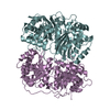
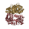
- Assembly
| Deposited unit | 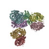
| ||||||||
|---|---|---|---|---|---|---|---|---|---|
| 1 | 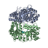
| ||||||||
| 2 | 
| ||||||||
| 3 | 
| ||||||||
| 4 | 
| ||||||||
| 5 | 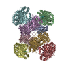
| ||||||||
| Unit cell |
|
-Components
-MITOCHONDRIAL PROCESSING PEPTIDASE ... , 2 types, 8 molecules ACEGBDFH
| #1: Protein | Mass: 52532.387 Da / Num. of mol.: 4 Source method: isolated from a genetically manipulated source Source: (gene. exp.) Plasmid: PTRC99A / Species (production host): Escherichia coli / Production host:  References: UniProt: P11914, mitochondrial processing peptidase #2: Protein | Mass: 48894.039 Da / Num. of mol.: 4 / Mutation: E73Q Source method: isolated from a genetically manipulated source Source: (gene. exp.) Plasmid: PTRC99A / Species (production host): Escherichia coli / Production host: References: UniProt: P10507, mitochondrial processing peptidase |
|---|
-Protein/peptide , 1 types, 4 molecules OPQR
| #3: Protein/peptide | Mass: 2862.421 Da / Num. of mol.: 4 / Fragment: RESIDUES 2-25 / Source method: obtained synthetically Details: The 24 residue peptide of CYTOCHROME C OXIDASE POLYPEPTIDE IV was synthesized using standard solid-phase Fmoc synthesis methodology References: UniProt: P04037, cytochrome-c oxidase |
|---|
-Non-polymers , 3 types, 10 molecules 


| #4: Chemical |  Mass: 238.305 Da / Num. of mol.: 2 / Source method: obtained synthetically / Formula: C8H18N2O4S / Comment: pH buffer*YM Mass: 238.305 Da / Num. of mol.: 2 / Source method: obtained synthetically / Formula: C8H18N2O4S / Comment: pH buffer*YM#5: Chemical | ChemComp-ZN /  Mass: 65.409 Da / Num. of mol.: 4 / Source method: obtained synthetically / Formula: Zn Mass: 65.409 Da / Num. of mol.: 4 / Source method: obtained synthetically / Formula: Zn#6: Water | ChemComp-HOH / | Mass: 18.015 Da / Num. of mol.: 4 / Source method: isolated from a natural source / Formula: H2O |
|---|
-Experimental details
-Experiment
| Experiment | Method: X-RAY DIFFRACTION / Number of used crystals: 1 |
|---|
- Sample preparation
| Crystal | Density Matthews: 3 Å3/Da / Density % sol: 59 % | ||||||||||||||||||||||||||||||||||||||||||||||||||||||||||||
|---|---|---|---|---|---|---|---|---|---|---|---|---|---|---|---|---|---|---|---|---|---|---|---|---|---|---|---|---|---|---|---|---|---|---|---|---|---|---|---|---|---|---|---|---|---|---|---|---|---|---|---|---|---|---|---|---|---|---|---|---|---|
| Crystal grow | *PLUS Temperature: 21 ℃ / pH: 7.5 / Method: vapor diffusion, hanging drop | ||||||||||||||||||||||||||||||||||||||||||||||||||||||||||||
| Components of the solutions | *PLUS
|
-Data collection
| Diffraction | Mean temperature: 105 K |
|---|---|
| Diffraction source | Source: SYNCHROTRON / Site: CHESS  / Beamline: F1 / Wavelength: 0.943 Å / Beamline: F1 / Wavelength: 0.943 Å |
| Detector | Type: ADSC QUANTUM 4 / Detector: CCD / Date: Jul 2, 2000 |
| Radiation | Protocol: SINGLE WAVELENGTH / Monochromatic (M) / Laue (L): M / Scattering type: x-ray |
| Radiation wavelength | Wavelength: 0.943 Å / Relative weight: 1 |
| Reflection | Resolution: 2.7→50 Å / Num. all: 134234 / Num. obs: 134234 / % possible obs: 99.9 % / Redundancy: 6.1 % / Biso Wilson estimate: 47.1 Å2 / Rmerge(I) obs: 0.079 / Net I/σ(I): 23.4 |
| Reflection shell | Resolution: 2.7→2.8 Å / Redundancy: 6.1 % / Rmerge(I) obs: 0.543 / Mean I/σ(I) obs: 3.5 / % possible all: 100 |
| Reflection | *PLUS Num. measured all: 820094 |
| Reflection shell | *PLUS % possible obs: 100 % |
- Processing
| Software |
| ||||||||||||||||||||||||||||||||||||||||||||||||||||||||||||
|---|---|---|---|---|---|---|---|---|---|---|---|---|---|---|---|---|---|---|---|---|---|---|---|---|---|---|---|---|---|---|---|---|---|---|---|---|---|---|---|---|---|---|---|---|---|---|---|---|---|---|---|---|---|---|---|---|---|---|---|---|---|
| Refinement | Starting model: PDB ID 1HR6 Resolution: 2.7→48.63 Å / Rfactor Rfree error: 0.006 / Data cutoff high absF: 6298765.76 / Data cutoff low absF: 0 / Isotropic thermal model: GROUP / Cross valid method: THROUGHOUT / σ(F): 0 / Stereochemistry target values: Maximum Likelihood Function
| ||||||||||||||||||||||||||||||||||||||||||||||||||||||||||||
| Solvent computation | Solvent model: FLAT MODEL / Bsol: 29.8593 Å2 / ksol: 0.323 e/Å3 | ||||||||||||||||||||||||||||||||||||||||||||||||||||||||||||
| Displacement parameters | Biso mean: 58.7 Å2
| ||||||||||||||||||||||||||||||||||||||||||||||||||||||||||||
| Refine analyze |
| ||||||||||||||||||||||||||||||||||||||||||||||||||||||||||||
| Refinement step | Cycle: LAST / Resolution: 2.7→48.63 Å
| ||||||||||||||||||||||||||||||||||||||||||||||||||||||||||||
| Refine LS restraints |
| ||||||||||||||||||||||||||||||||||||||||||||||||||||||||||||
| Refine LS restraints NCS | NCS model details: RESTRAINED | ||||||||||||||||||||||||||||||||||||||||||||||||||||||||||||
| LS refinement shell | Resolution: 2.7→2.8 Å / Rfactor Rfree error: 0.024 / Total num. of bins used: 10
| ||||||||||||||||||||||||||||||||||||||||||||||||||||||||||||
| Xplor file |
| ||||||||||||||||||||||||||||||||||||||||||||||||||||||||||||
| Software | *PLUS Name: CNS / Version: 1 / Classification: refinement | ||||||||||||||||||||||||||||||||||||||||||||||||||||||||||||
| Refinement | *PLUS σ(F): 0 / % reflection Rfree: 1.5 % | ||||||||||||||||||||||||||||||||||||||||||||||||||||||||||||
| Solvent computation | *PLUS | ||||||||||||||||||||||||||||||||||||||||||||||||||||||||||||
| Displacement parameters | *PLUS Biso mean: 58.7 Å2 | ||||||||||||||||||||||||||||||||||||||||||||||||||||||||||||
| Refine LS restraints | *PLUS
| ||||||||||||||||||||||||||||||||||||||||||||||||||||||||||||
| LS refinement shell | *PLUS Rfactor Rfree: 0.328 / % reflection Rfree: 1.4 % / Rfactor Rwork: 0.303 |
