 Movie
Movie Controller

[English] 日本語
 Yorodumi
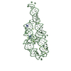
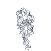
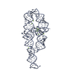
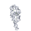
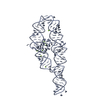
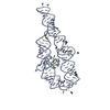
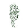
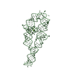
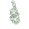
- PDB-1hr2: CRYSTAL STRUCTURE ANALYSIS OF A MUTANT P4-P6 DOMAIN (DELC209) OF ... -
+ Open data

- Basic information
| Entry | Database: PDB / ID: 1hr2 | ||||||
|---|---|---|---|---|---|---|---|
| Title | CRYSTAL STRUCTURE ANALYSIS OF A MUTANT P4-P6 DOMAIN (DELC209) OF TETRAHYMENA THEMOPHILA GROUP I INTRON. | ||||||
 Components Components | P4-P6 DELC209 MUTANT RNA RIBOZYME DOMAIN | ||||||
 Keywords Keywords | RNA / P4-P6 / C209 / RIBOZYME / TETRAHYMENA / GROUP I INTRON / RIBONUCLEIC ACID | ||||||
| Function / homology | RNA / RNA (> 10) / RNA (> 100) Function and homology information Function and homology information | ||||||
| Method |  X-RAY DIFFRACTION / SYNCHROTRON / MOLECULAR REPLACEMENT / Resolution: 2.25 Å X-RAY DIFFRACTION / SYNCHROTRON / MOLECULAR REPLACEMENT / Resolution: 2.25 Å | ||||||
 Authors Authors | Juneau, K. / Podell, E.R. / Harrington, D.J. / Cech, T.R. | ||||||
 Citation Citation | Journal: Structure / Year: 2001 Title: Structural basis of the enhanced stability of a mutant ribozyme domain and a detailed view of RNA--solvent interactions. Authors: Juneau, K. / Podell, E. / Harrington, D.J. / Cech, T.R. #1: Journal: RNA / Year: 1999Title: In-Vitro Selection of RNAs With Increased Tertiary Structure Stability Authors: Juneau, K. / Cech, T.R. #2: Journal: Science / Year: 1996Title: Crystal Structure of a Group I Ribozyme Domain: Principles of RNA Packing Authors: Cate, J.H. / Gooding, A.R. / Podell, E. / Zhou, K. / Golden, B.L. / Kundrot, C.E. / Cech, T.R. / A Doudna, J. #3: Journal: Structure / Year: 1996Title: Metal-binding Sites in the Major Groove of a Large Ribozyme Domain Authors: Cate, J.H. / Doudna, J.A. #4: Journal: Nat.Struct.Biol. / Year: 1997Title: A Magnesium Ion Core at the Heart of a Ribozyme Domain Authors: Cate, J.H. / Hanna, R.L. / Doudna, J.A. | ||||||
| History |
|
- Structure visualization
| Structure viewer | Molecule: MolmilJmol/JSmol |
|---|
- Downloads & links
-Download
| PDBx/mmCIF format | 1hr2.cif.gz | 194.8 KB | Display | PDBx/mmCIF format |
|---|---|---|---|---|
| PDB format | pdb1hr2.ent.gz | 146.5 KB | Display | PDB format |
| PDBx/mmJSON format | 1hr2.json.gz | Tree view | PDBx/mmJSON format | |
| Others |  Other downloads Other downloads |
-Validation report
| Arichive directory | https://data.pdbj.org/pub/pdb/validation_reports/hr/1hr2ftp://data.pdbj.org/pub/pdb/validation_reports/hr/1hr2 | HTTPS FTP |
|---|
-Related structure data
| Related structure data | 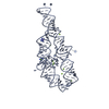 1gidS S: Starting model for refinement |
|---|---|
| Similar structure data |

-Links
 PDBj
PDBj


- Assembly
| Deposited unit | 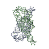
| ||||||||
|---|---|---|---|---|---|---|---|---|---|
| 1 | 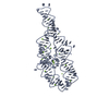
| ||||||||
| 2 | 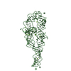
| ||||||||
| Unit cell |
|
-Components
| #1: RNA chain | Mass: 51091.352 Da / Num. of mol.: 2 / Mutation: DELETION OF C209 / Source method: obtained synthetically Details: The RNA was prepared by transcription with T7 RNA polymerase #2: Chemical | ChemComp-MG /   Mass: 24.305 Da / Num. of mol.: 42 / Source method: obtained synthetically / Formula: Mg Mass: 24.305 Da / Num. of mol.: 42 / Source method: obtained synthetically / Formula: Mg#3: Water | ChemComp-HOH / |  Mass: 18.015 Da / Num. of mol.: 265 / Source method: isolated from a natural source / Formula: H2O Mass: 18.015 Da / Num. of mol.: 265 / Source method: isolated from a natural source / Formula: H2O |
|---|
-Experimental details
-Experiment
| Experiment | Method: X-RAY DIFFRACTION / Number of used crystals: 1 |
|---|
- Sample preparation
| Crystal | Density Matthews: 3.37 Å3/Da / Density % sol: 63.46 % | ||||||||||||||||||||||||||||||||||||||||||||||||||||||||||||||||||||||||||||||||||||
|---|---|---|---|---|---|---|---|---|---|---|---|---|---|---|---|---|---|---|---|---|---|---|---|---|---|---|---|---|---|---|---|---|---|---|---|---|---|---|---|---|---|---|---|---|---|---|---|---|---|---|---|---|---|---|---|---|---|---|---|---|---|---|---|---|---|---|---|---|---|---|---|---|---|---|---|---|---|---|---|---|---|---|---|---|---|
| Crystal grow | Temperature: 293 K / Method: vapor diffusion, sitting drop / pH: 6.5 Details: MPD, magnesium chloride, sodium cacodylate, spermine, sodium chloride, pH 6.5, VAPOR DIFFUSION, SITTING DROP, temperature 293K | ||||||||||||||||||||||||||||||||||||||||||||||||||||||||||||||||||||||||||||||||||||
| Components of the solutions |
| ||||||||||||||||||||||||||||||||||||||||||||||||||||||||||||||||||||||||||||||||||||
| Crystal grow | *PLUS Temperature: 30 ℃Details: drop is set up using 2:8:5 ratio of solutions a, b and c | ||||||||||||||||||||||||||||||||||||||||||||||||||||||||||||||||||||||||||||||||||||
| Components of the solutions | *PLUS
|
-Data collection
| Diffraction | Mean temperature: 100 K |
|---|---|
| Diffraction source | Source: SYNCHROTRON / Site: ALS  / Beamline: 5.0.1 / Wavelength: 1.1 Å / Beamline: 5.0.1 / Wavelength: 1.1 Å |
| Detector | Type: ADSC QUANTUM 4 / Detector: CCD / Date: Mar 24, 1999 / Details: W16 wiggler |
| Radiation | Monochromator: single crystal, cylindrically bent / Protocol: SINGLE WAVELENGTH / Monochromatic (M) / Laue (L): M / Scattering type: x-ray |
| Radiation wavelength | Wavelength: 1.1 Å / Relative weight: 1 |
| Reflection | Resolution: 2.25→30 Å / Num. all: 64900 / Num. obs: 64900 / % possible obs: 98 % / Observed criterion σ(I): -3 / Redundancy: 4.8 % / Rmerge(I) obs: 0.051 / Net I/σ(I): 22.7 |
| Reflection shell | Resolution: 2.25→2.33 Å / Redundancy: 4.4 % / Rmerge(I) obs: 0.578 / Mean I/σ(I) obs: 2.4 / Num. unique all: 6463 / Rsym value: 57.8 / % possible all: 99.3 |
| Reflection shell | *PLUS % possible obs: 99.3 % |
- Processing
| Software |
| ||||||||||||||||||||||||||||||||||||||||||
|---|---|---|---|---|---|---|---|---|---|---|---|---|---|---|---|---|---|---|---|---|---|---|---|---|---|---|---|---|---|---|---|---|---|---|---|---|---|---|---|---|---|---|---|
| Refinement | Method to determine structure: MOLECULAR REPLACEMENT Starting model: PDB entry 1gid Resolution: 2.25→30 Å / Isotropic thermal model: isotropic / Cross valid method: THROUGHOUT / σ(F): 0 / σ(I): 0 Stereochemistry target values: G. Parkinson, J. Vojtechovsky, L. Clowney, A.T. Brunger, H.M. Berman, New Parameters for the Refinement of Nucleic Acid Containing Structures, Acta Cryst. D, 52, 57-64 (1996). Details: used maximum likelihood function as implemented by CNS
| ||||||||||||||||||||||||||||||||||||||||||
| Displacement parameters | Biso mean: 75.4 Å2
| ||||||||||||||||||||||||||||||||||||||||||
| Refine analyze |
| ||||||||||||||||||||||||||||||||||||||||||
| Refinement step | Cycle: LAST / Resolution: 2.25→30 Å
| ||||||||||||||||||||||||||||||||||||||||||
| Refine LS restraints |
| ||||||||||||||||||||||||||||||||||||||||||
| LS refinement shell |
| ||||||||||||||||||||||||||||||||||||||||||
| Software | *PLUS Name: CNS / Classification: refinement | ||||||||||||||||||||||||||||||||||||||||||
| Refine LS restraints | *PLUS
|
