 Movie
Movie Controller

[English] 日本語
 Yorodumi
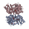
- PDB-1hor: STRUCTURE AND CATALYTIC MECHANISM OF GLUCOSAMINE 6-PHOSPHATE DEAM... -
+ Open data

- Basic information
| Entry | Database: PDB / ID: 1hor | ||||||
|---|---|---|---|---|---|---|---|
| Title | STRUCTURE AND CATALYTIC MECHANISM OF GLUCOSAMINE 6-PHOSPHATE DEAMINASE FROM ESCHERICHIA COLI AT 2.1 ANGSTROMS RESOLUTION | ||||||
 Components Components | GLUCOSAMINE 6-PHOSPHATE DEAMINASE | ||||||
 Keywords Keywords | INTRAMOLECULAR OXIDOREDUCTASE DEAMINASE | ||||||
| Function / homology |  Function and homology information Function and homology informationD-glucosamine catabolic process / glucosamine-6-phosphate deaminase / glucosamine-6-phosphate deaminase activity / N-acetylglucosamine catabolic process / N-acetylneuraminate catabolic process / UDP-N-acetylglucosamine biosynthetic process / carbohydrate metabolic process / identical protein binding / cytosol / cytoplasm Similarity search - Function | ||||||
| Biological species |  | ||||||
| Method |  X-RAY DIFFRACTION / Resolution: 2.4 Å X-RAY DIFFRACTION / Resolution: 2.4 Å | ||||||
 Authors Authors | Oliva, G. / Fontes, M.R.M. / Garratt, R.C. / Altamirano, M.M. / Calcagno, M.L. / Horjales, E. | ||||||
 Citation Citation | Journal: Structure / Year: 1995 Title: Structure and catalytic mechanism of glucosamine 6-phosphate deaminase from Escherichia coli at 2.1 A resolution. Authors: Oliva, G. / Fontes, M.R. / Garratt, R.C. / Altamirano, M.M. / Calcagno, M.L. / Horjales, E. | ||||||
| History |
|
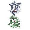
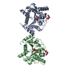
- Structure visualization
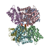
| Structure viewer | Molecule: MolmilJmol/JSmol |
|---|
- Downloads & links
-Download
| PDBx/mmCIF format | 1hor.cif.gz | 117.8 KB | Display | PDBx/mmCIF format |
|---|---|---|---|---|
| PDB format | pdb1hor.ent.gz | 93.2 KB | Display | PDB format |
| PDBx/mmJSON format | 1hor.json.gz | Tree view | PDBx/mmJSON format | |
| Others |  Other downloads Other downloads |
-Validation report
| Arichive directory | https://data.pdbj.org/pub/pdb/validation_reports/ho/1horftp://data.pdbj.org/pub/pdb/validation_reports/ho/1hor | HTTPS FTP |
|---|
-Related structure data


-Links
 PDBj
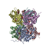
PDBj- Assembly
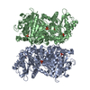
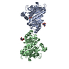
| Deposited unit | 
| ||||||||
|---|---|---|---|---|---|---|---|---|---|
| 1 | 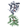
| ||||||||
| Unit cell |
|
-Components
| #1: Protein | Mass: 29812.211 Da / Num. of mol.: 2 Source method: isolated from a genetically manipulated source Source: (gene. exp.) #2: Chemical |   Mass: 94.971 Da / Num. of mol.: 2 / Source method: obtained synthetically / Formula: PO4 Mass: 94.971 Da / Num. of mol.: 2 / Source method: obtained synthetically / Formula: PO4#3: Chemical | 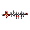  Mass: 261.167 Da / Num. of mol.: 2 / Source method: obtained synthetically / Formula: C6H16NO8P Mass: 261.167 Da / Num. of mol.: 2 / Source method: obtained synthetically / Formula: C6H16NO8P#4: Water | ChemComp-HOH / |  Mass: 18.015 Da / Num. of mol.: 216 / Source method: isolated from a natural source / Formula: H2O Mass: 18.015 Da / Num. of mol.: 216 / Source method: isolated from a natural source / Formula: H2O |
|---|
-Experimental details
-Experiment
| Experiment | Method: X-RAY DIFFRACTION |
|---|
- Sample preparation
| Crystal | Density Matthews: 2.84 Å3/Da / Density % sol: 56.7 % | |||||||||||||||
|---|---|---|---|---|---|---|---|---|---|---|---|---|---|---|---|---|
| Crystal grow | *PLUS Temperature: 20 ℃ / pH: 8.2 / Method: vapor diffusion, hanging drop | |||||||||||||||
| Components of the solutions | *PLUS
|
-Data collection
| Radiation | Scattering type: x-ray |
|---|---|
| Radiation wavelength | Relative weight: 1 |
| Reflection | *PLUS Num. obs: 18437 / % possible obs: 81.1 % / Rmerge(I) obs: 0.069 |
- Processing
| Software |
| ||||||||||||||||||||||||||||||||||||||||||||||||||||||||||||
|---|---|---|---|---|---|---|---|---|---|---|---|---|---|---|---|---|---|---|---|---|---|---|---|---|---|---|---|---|---|---|---|---|---|---|---|---|---|---|---|---|---|---|---|---|---|---|---|---|---|---|---|---|---|---|---|---|---|---|---|---|---|
| Refinement | Resolution: 2.4→8 Å / Rfactor Rwork: 0.165 / Rfactor obs: 0.165 | ||||||||||||||||||||||||||||||||||||||||||||||||||||||||||||
| Refinement step | Cycle: LAST / Resolution: 2.4→8 Å
| ||||||||||||||||||||||||||||||||||||||||||||||||||||||||||||
| Refine LS restraints |
| ||||||||||||||||||||||||||||||||||||||||||||||||||||||||||||
| Refinement | *PLUS Rfactor Rfree: 0.225 | ||||||||||||||||||||||||||||||||||||||||||||||||||||||||||||
| Solvent computation | *PLUS | ||||||||||||||||||||||||||||||||||||||||||||||||||||||||||||
| Displacement parameters | *PLUS | ||||||||||||||||||||||||||||||||||||||||||||||||||||||||||||
| Refine LS restraints | *PLUS
|
