 Movie
Movie Controller

[English] 日本語
 Yorodumi
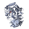
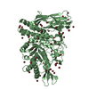
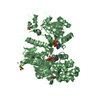
- PDB-1ho5: 5'-NUCLEOTIDASE (E. COLI) IN COMPLEX WITH ADENOSINE AND PHOSPHATE -
+ Open data

- Basic information
| Entry | Database: PDB / ID: 1ho5 | ||||||
|---|---|---|---|---|---|---|---|
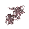
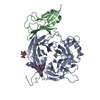
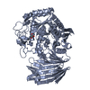
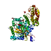
| Title | 5'-NUCLEOTIDASE (E. COLI) IN COMPLEX WITH ADENOSINE AND PHOSPHATE | ||||||
 Components Components | 5'-NUCLEOTIDASE | ||||||
 Keywords Keywords | HYDROLASE / metalloprotein / domain movement | ||||||
| Function / homology |  Function and homology information Function and homology informationUDP-sugar diphosphatase / UDP-sugar diphosphatase activity / nucleotide catabolic process / 5'-nucleotidase / 5'-nucleotidase activity / outer membrane-bounded periplasmic space / nucleotide binding / metal ion binding Similarity search - Function | ||||||
| Biological species |  | ||||||
| Method |  X-RAY DIFFRACTION / SYNCHROTRON / MOLECULAR REPLACEMENT / Resolution: 2.1 Å X-RAY DIFFRACTION / SYNCHROTRON / MOLECULAR REPLACEMENT / Resolution: 2.1 Å | ||||||
 Authors Authors | Knoefel, T. / Straeter, N. | ||||||
 Citation Citation | Journal: J.Mol.Biol. / Year: 2001 Title: Mechanism of hydrolysis of phosphate esters by the dimetal center of 5'-nucleotidase based on crystal structures. Authors: Knofel, T. / Strater, N. #1: Journal: J.Mol.Biol. / Year: 2001Title: E. COLI 5'-NUCLEOTIDASE UNDERGOES A HINGE-BENDING DOMAIN ROTATION RESEMBLING A BALL-AND-SOCKET MOTION Authors: Knoefel, T. / Straeter, N. #2: Journal: Nat.Struct.Biol. / Year: 1999Title: X-ray structure of the Escherichia coli periplasmic 5'-nucleotidase containing a dimetal catalytic site Authors: Knoefel, T. / Straeter, N. | ||||||
| History |
|
- Structure visualization
| Structure viewer | Molecule: MolmilJmol/JSmol |
|---|
- Downloads & links
-Download
| PDBx/mmCIF format | 1ho5.cif.gz | 224.9 KB | Display | PDBx/mmCIF format |
|---|---|---|---|---|
| PDB format | pdb1ho5.ent.gz | 176.6 KB | Display | PDB format |
| PDBx/mmJSON format | 1ho5.json.gz | Tree view | PDBx/mmJSON format | |
| Others |  Other downloads Other downloads |
-Validation report
| Arichive directory | https://data.pdbj.org/pub/pdb/validation_reports/ho/1ho5ftp://data.pdbj.org/pub/pdb/validation_reports/ho/1ho5 | HTTPS FTP |
|---|
-Related structure data
| Related structure data |  1hp1C  1hpuC  1ushS S: Starting model for refinement C: citing same article ( |
|---|---|
| Similar structure data |


-Links
 PDBj
PDBj

- Assembly
| Deposited unit | 
| ||||||||
|---|---|---|---|---|---|---|---|---|---|
| 1 | 
| ||||||||
| 2 | 
| ||||||||
| Unit cell |
|
-Components
| #1: Protein | Mass: 58277.598 Da / Num. of mol.: 2 Source method: isolated from a genetically manipulated source Source: (gene. exp.) References: UniProt: P07024, 5'-nucleotidase, UDP-sugar diphosphatase #2: Chemical | ChemComp-MN /   Mass: 54.938 Da / Num. of mol.: 4 / Source method: obtained synthetically / Formula: Mn Mass: 54.938 Da / Num. of mol.: 4 / Source method: obtained synthetically / Formula: Mn#3: Chemical |   Mass: 94.971 Da / Num. of mol.: 2 / Source method: obtained synthetically / Formula: PO4 Mass: 94.971 Da / Num. of mol.: 2 / Source method: obtained synthetically / Formula: PO4#4: Chemical |   Mass: 267.241 Da / Num. of mol.: 2 / Source method: obtained synthetically / Formula: C10H13N5O4 Mass: 267.241 Da / Num. of mol.: 2 / Source method: obtained synthetically / Formula: C10H13N5O4#5: Water | ChemComp-HOH / |  Mass: 18.015 Da / Num. of mol.: 355 / Source method: isolated from a natural source / Formula: H2O Mass: 18.015 Da / Num. of mol.: 355 / Source method: isolated from a natural source / Formula: H2OHas protein modification | Y | |
|---|
-Experimental details
-Experiment
| Experiment | Method: X-RAY DIFFRACTION / Number of used crystals: 1 |
|---|
- Sample preparation
| Crystal | Density Matthews: 2.52 Å3/Da / Density % sol: 51.09 % | ||||||||||||||||||||||||||||||||||||||||||||||||||||||||
|---|---|---|---|---|---|---|---|---|---|---|---|---|---|---|---|---|---|---|---|---|---|---|---|---|---|---|---|---|---|---|---|---|---|---|---|---|---|---|---|---|---|---|---|---|---|---|---|---|---|---|---|---|---|---|---|---|---|
| Crystal grow | Temperature: 298 K / Method: vapor diffusion, hanging drop / pH: 6 Details: sodium citrate, lithium chloride, potassium phosphate, adenosine, manganese chloride, PEG monomethylether 5000, pH 6.0, VAPOR DIFFUSION, HANGING DROP, temperature 298.0K | ||||||||||||||||||||||||||||||||||||||||||||||||||||||||
| Crystal grow | *PLUS Method: vapor diffusion / Details: Knofel, T., (2001) J.Mol.Biol., 309, 255. | ||||||||||||||||||||||||||||||||||||||||||||||||||||||||
| Components of the solutions | *PLUS
|
-Data collection
| Diffraction | Mean temperature: 100 K |
|---|---|
| Diffraction source | Source: SYNCHROTRON / Site: EMBL/DESY, HAMBURG  / Beamline: X11 / Wavelength: 0.9116 Å / Beamline: X11 / Wavelength: 0.9116 Å |
| Detector | Type: MARRESEARCH / Detector: IMAGE PLATE / Date: Mar 25, 1999 |
| Radiation | Monochromator: bent single-crystal germanium triangular monochromator Protocol: SINGLE WAVELENGTH / Monochromatic (M) / Laue (L): M / Scattering type: x-ray |
| Radiation wavelength | Wavelength: 0.9116 Å / Relative weight: 1 |
| Reflection | Resolution: 2→50 Å / Num. all: 69235 / Num. obs: 69235 / % possible obs: 99.5 % / Observed criterion σ(F): 0 / Observed criterion σ(I): 0 / Redundancy: 3.5 % / Biso Wilson estimate: 20.4 Å2 / Rmerge(I) obs: 0.054 / Net I/σ(I): 13.1 |
| Reflection shell | Resolution: 2.1→2.2 Å / Redundancy: 3.1 % / Rmerge(I) obs: 0.298 / % possible all: 97.2 |
| Reflection | *PLUS |
- Processing
| Software |
| ||||||||||||||||||||||||||||||||||||||||||||
|---|---|---|---|---|---|---|---|---|---|---|---|---|---|---|---|---|---|---|---|---|---|---|---|---|---|---|---|---|---|---|---|---|---|---|---|---|---|---|---|---|---|---|---|---|---|
| Refinement | Method to determine structure: MOLECULAR REPLACEMENT Starting model: 1ush Resolution: 2.1→29.56 Å / Rfactor Rfree error: 0.006 / Data cutoff high absF: 2757655.82 / Data cutoff low absF: 0 / Isotropic thermal model: RESTRAINED / Cross valid method: THROUGHOUT / σ(F): 0 / σ(I): 0 / Stereochemistry target values: Engh & Huber
| ||||||||||||||||||||||||||||||||||||||||||||
| Solvent computation | Solvent model: FLAT MODEL / Bsol: 41.2 Å2 / ksol: 0.3247 e/Å3 | ||||||||||||||||||||||||||||||||||||||||||||
| Displacement parameters | Biso mean: 45.3 Å2
| ||||||||||||||||||||||||||||||||||||||||||||
| Refine analyze |
| ||||||||||||||||||||||||||||||||||||||||||||
| Refinement step | Cycle: LAST / Resolution: 2.1→29.56 Å
| ||||||||||||||||||||||||||||||||||||||||||||
| Refine LS restraints |
| ||||||||||||||||||||||||||||||||||||||||||||
| LS refinement shell | Resolution: 2.1→2.23 Å / Rfactor Rfree error: 0.017 / Total num. of bins used: 6
| ||||||||||||||||||||||||||||||||||||||||||||
| Refinement | *PLUS | ||||||||||||||||||||||||||||||||||||||||||||
| Solvent computation | *PLUS | ||||||||||||||||||||||||||||||||||||||||||||
| Displacement parameters | *PLUS | ||||||||||||||||||||||||||||||||||||||||||||
| Refine LS restraints | *PLUS
| ||||||||||||||||||||||||||||||||||||||||||||
| LS refinement shell | *PLUS Rfactor Rfree: 0.324 / Rfactor Rwork: 0.269 |
