 Movie
Movie Controller


+ Open data

- Basic information
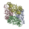
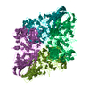
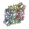
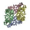
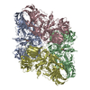
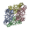
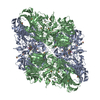
| Entry | Database: PDB / ID: 1hn1 | ||||||
|---|---|---|---|---|---|---|---|
| Title | E. COLI (LAC Z) BETA-GALACTOSIDASE (ORTHORHOMBIC) | ||||||
 Components Components | BETA-GALACTOSIDASE | ||||||
 Keywords Keywords | HYDROLASE / ALPHA/BETA BARREL / JELLY ROLL BARREL / FIBRONECTIN / BETA SUPERSANDWICH | ||||||
| Function / homology |  Function and homology information Function and homology informationalkali metal ion binding / lactose catabolic process / beta-galactosidase complex / beta-galactosidase / beta-galactosidase activity / carbohydrate binding / magnesium ion binding / identical protein binding Similarity search - Function | ||||||
| Biological species |  | ||||||
| Method |  X-RAY DIFFRACTION / MOLECULAR REPLACEMENT / Resolution: 3 Å X-RAY DIFFRACTION / MOLECULAR REPLACEMENT / Resolution: 3 Å | ||||||
 Authors Authors | Juers, D.H. / Matthews, B.W. | ||||||
 Citation Citation | Journal: J.Mol.Biol. / Year: 2001 Title: Reversible lattice repacking illustrates the temperature dependence of macromolecular interactions. Authors: Juers, D.H. / Matthews, B.W. #1: Journal: Protein Sci. / Year: 2000Title: High resolution refinement of beta-galactosidase in a new crystal form reveals multiple metal-binding sites and provides a structural basis for alpha-complementation Authors: JUERS, D.H. / JACOBSON, R.H. / WIGLEY, D. / ZHANG, X.J. / HUBER, R.E. / TRONRUD, D.E. / MATTHEWS, B.W. #2: Journal: Protein Sci. / Year: 1999Title: Structural comparisons of TIM barrel proteins suggest functional and evolutionary relationships between beta-galactosidase and other glycohydrolases Authors: JUERS, D.H. / HUBER, R.E. / MATTHEWS, B.W. #3: Journal: Nature / Year: 1994Title: Three-dimensional structure of beta-galactosidase from E. coli. Authors: JACOBSON, R.H. / ZHANG, X.J. / DUBOSE, R.F. / MATTHEWS, B.W. #4: Journal: J.Mol.Biol. / Year: 1992Title: Crystallization of beta-galactosidase from Escherichia coli. Authors: JACOBSON, R.H. / MATTHEWS, B.W. | ||||||
| History |
|
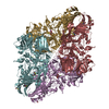
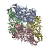
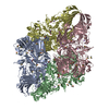
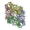
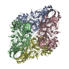
- Structure visualization
| Structure viewer | Molecule: MolmilJmol/JSmol |
|---|
- Downloads & links
-Download
| PDBx/mmCIF format | 1hn1.cif.gz | 822.2 KB | Display | PDBx/mmCIF format |
|---|---|---|---|---|
| PDB format | pdb1hn1.ent.gz | 664.4 KB | Display | PDB format |
| PDBx/mmJSON format | 1hn1.json.gz | Tree view | PDBx/mmJSON format | |
| Others |  Other downloads Other downloads |
-Validation report
| Arichive directory | https://data.pdbj.org/pub/pdb/validation_reports/hn/1hn1ftp://data.pdbj.org/pub/pdb/validation_reports/hn/1hn1 | HTTPS FTP |
|---|
-Related structure data
| Related structure data | 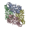 1dp0S S: Starting model for refinement |
|---|---|
| Similar structure data |


-Links
 PDBj
PDBj


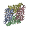
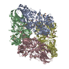
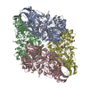
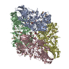
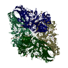
- Assembly
| Deposited unit | 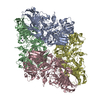
| ||||||||
|---|---|---|---|---|---|---|---|---|---|
| 1 |
| ||||||||
| Unit cell |
|
-Components
| #1: Protein | Mass: 116506.266 Da / Num. of mol.: 4 Source method: isolated from a genetically manipulated source Source: (gene. exp.) References: UniProt: P00722, 4-hydroxybenzoyl-CoA thioesterase #2: Chemical | ChemComp-MG /   Mass: 24.305 Da / Num. of mol.: 8 / Source method: obtained synthetically / Formula: Mg Mass: 24.305 Da / Num. of mol.: 8 / Source method: obtained synthetically / Formula: Mg#3: Chemical | ChemComp-NA /   Mass: 22.990 Da / Num. of mol.: 7 / Source method: obtained synthetically / Formula: Na Mass: 22.990 Da / Num. of mol.: 7 / Source method: obtained synthetically / Formula: Na#4: Water | ChemComp-HOH / |  Mass: 18.015 Da / Num. of mol.: 401 / Source method: isolated from a natural source / Formula: H2O Mass: 18.015 Da / Num. of mol.: 401 / Source method: isolated from a natural source / Formula: H2O |
|---|
-Experimental details
-Experiment
| Experiment | Method: X-RAY DIFFRACTION / Number of used crystals: 2 |
|---|
- Sample preparation
| Crystal | Density Matthews: 2.89 Å3/Da / Density % sol: 57.47 % | ||||||||||||||||||||||||||||||||||||||||||
|---|---|---|---|---|---|---|---|---|---|---|---|---|---|---|---|---|---|---|---|---|---|---|---|---|---|---|---|---|---|---|---|---|---|---|---|---|---|---|---|---|---|---|---|
| Crystal grow | Temperature: 288 K / Method: vapor diffusion, hanging drop / pH: 6.5 Details: 10% PEG 8000, 100 mM BIS TRIS, 200 mM MGCL(2), 100 mM NACL, 10 mM DTT, pH 6.5, VAPOR DIFFUSION, HANGING DROP, temperature 288K | ||||||||||||||||||||||||||||||||||||||||||
| Crystal grow | *PLUS Method: vapor diffusionDetails: Juers, D.H., (2000) Protein Sci., 9, 1685., used seeding | ||||||||||||||||||||||||||||||||||||||||||
| Components of the solutions | *PLUS
|
-Data collection
| Diffraction | Mean temperature: 298 K |
|---|---|
| Diffraction source | Source: ROTATING ANODE / Type: RIGAKU / Wavelength: 1.54 Å |
| Detector | Type: RIGAKU RAXIS IV / Detector: IMAGE PLATE / Date: Aug 1, 1999 / Details: mirrors |
| Radiation | Protocol: SINGLE WAVELENGTH / Monochromatic (M) / Laue (L): M / Scattering type: x-ray |
| Radiation wavelength | Wavelength: 1.54 Å / Relative weight: 1 |
| Reflection | Resolution: 3→23 Å / Num. all: 103758 / Num. obs: 103758 / % possible obs: 95.1 % / Observed criterion σ(F): 0 / Observed criterion σ(I): 0 / Redundancy: 2.2 % / Rmerge(I) obs: 0.092 / Net I/σ(I): 7.2 |
| Reflection shell | Highest resolution: 3 Å / Rmerge(I) obs: 0.292 / Mean I/σ(I) obs: 2.3 / % possible all: 98.5 |
- Processing
| Software |
| |||||||||||||||||||||||||||
|---|---|---|---|---|---|---|---|---|---|---|---|---|---|---|---|---|---|---|---|---|---|---|---|---|---|---|---|---|
| Refinement | Method to determine structure: MOLECULAR REPLACEMENT Starting model: PDB ENTRY 1DP0 Resolution: 3→15 Å / σ(F): 0 / σ(I): 0 / Stereochemistry target values: TNT
| |||||||||||||||||||||||||||
| Solvent computation | Solvent model: BABINET'S PRINCIPLE / Bsol: 200 Å2 / ksol: 0.75 e/Å3 | |||||||||||||||||||||||||||
| Displacement parameters | Biso mean: -0.3 Å2
| |||||||||||||||||||||||||||
| Refinement step | Cycle: LAST / Resolution: 3→15 Å
| |||||||||||||||||||||||||||
| Refine LS restraints |
|
