[English] 日本語
 Yorodumi
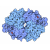
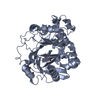
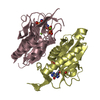
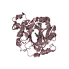
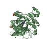
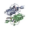
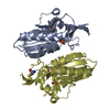
- PDB-1hgx: HYPOXANTHINE-GUANINE-XANTHINE PHOSPHORIBOSYLTRANSFERASE (HGXPRTASE) -
+ Open data
- Basic information
| Entry | Database: PDB / ID: 1hgx | ||||||
|---|---|---|---|---|---|---|---|
| Title | HYPOXANTHINE-GUANINE-XANTHINE PHOSPHORIBOSYLTRANSFERASE (HGXPRTASE) | ||||||
 Components Components | HYPOXANTHINE-GUANINE-XANTHINE PHOSPHORIBOSYLTRANSFERASE | ||||||
 Keywords Keywords | TRANSFERASE (GLYCOSYLTRANSFERASE) / TRANSFERASE / GLYCOSYLTRANSFERASE / PURINE SALVAGE | ||||||
| Function / homology |  Function and homology information Function and homology informationxanthine phosphoribosyltransferase / XMP salvage / xanthine phosphoribosyltransferase activity / hypoxanthine phosphoribosyltransferase / guanine phosphoribosyltransferase activity / guanine salvage / hypoxanthine metabolic process / hypoxanthine phosphoribosyltransferase activity / GMP salvage / IMP salvage ...xanthine phosphoribosyltransferase / XMP salvage / xanthine phosphoribosyltransferase activity / hypoxanthine phosphoribosyltransferase / guanine phosphoribosyltransferase activity / guanine salvage / hypoxanthine metabolic process / hypoxanthine phosphoribosyltransferase activity / GMP salvage / IMP salvage / purine ribonucleoside salvage / nucleotide binding / magnesium ion binding / cytosol Similarity search - Function | ||||||
| Biological species |  Tritrichomonas foetus (eukaryote) Tritrichomonas foetus (eukaryote) | ||||||
| Method |  X-RAY DIFFRACTION / X-RAY DIFFRACTION /  MOLECULAR REPLACEMENT / Resolution: 1.9 Å MOLECULAR REPLACEMENT / Resolution: 1.9 Å | ||||||
 Authors Authors | Somoza, J.R. / Wang, C.C. / Fletterick, R.J. | ||||||
 Citation Citation |  Journal: Biochemistry / Year: 1996 Journal: Biochemistry / Year: 1996Title: Crystal structure of the hypoxanthine-guanine-xanthine phosphoribosyltransferase from the protozoan parasite Tritrichomonas foetus. Authors: Somoza, J.R. / Chin, M.S. / Focia, P.J. / Wang, C.C. / Fletterick, R.J. #1:  Journal: Mol.Biochem.Parasitol. / Year: 1994 Journal: Mol.Biochem.Parasitol. / Year: 1994Title: Isolation, Sequencing and Expression of the Gene Encoding Hypoxanthine-Guanine-Xanthine Phosphoribosyltransferase of Tritrichomonas Foetus Authors: Chin, M.S. / Wang, C.C. | ||||||
| History |
|
- Structure visualization
| Structure viewer | Molecule:  Molmil Molmil Jmol/JSmol Jmol/JSmol |
|---|
- Downloads & links
- Download
| PDBx/mmCIF format |  1hgx.cif.gz 1hgx.cif.gz | 80.8 KB | Display |  PDBx/mmCIF format PDBx/mmCIF format |
|---|---|---|---|---|
| PDB format |  pdb1hgx.ent.gz pdb1hgx.ent.gz | 60.5 KB | Display |  PDB format PDB format |
| PDBx/mmJSON format |  1hgx.json.gz 1hgx.json.gz | Tree view |  PDBx/mmJSON format PDBx/mmJSON format | |
| Others |  Other downloads Other downloads |
-Validation report
| Summary document |  1hgx_validation.pdf.gz 1hgx_validation.pdf.gz | 750.1 KB | Display |  wwPDB validaton report wwPDB validaton report |
|---|---|---|---|---|
| Full document |  1hgx_full_validation.pdf.gz 1hgx_full_validation.pdf.gz | 751.8 KB | Display | |
| Data in XML |  1hgx_validation.xml.gz 1hgx_validation.xml.gz | 15.3 KB | Display | |
| Data in CIF |  1hgx_validation.cif.gz 1hgx_validation.cif.gz | 21.2 KB | Display | |
| Arichive directory |  https://data.pdbj.org/pub/pdb/validation_reports/hg/1hgx https://data.pdbj.org/pub/pdb/validation_reports/hg/1hgx ftp://data.pdbj.org/pub/pdb/validation_reports/hg/1hgx ftp://data.pdbj.org/pub/pdb/validation_reports/hg/1hgx | HTTPS FTP |
-Related structure data
| Similar structure data |
|---|
- Links
Links
- Assembly
| Deposited unit | 
| ||||||||
|---|---|---|---|---|---|---|---|---|---|
| 1 |
| ||||||||
| Unit cell |
|
- Components
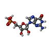
| #1: Protein | Mass: 21114.301 Da / Num. of mol.: 2 Source method: isolated from a genetically manipulated source Source: (gene. exp.)  Tritrichomonas foetus (eukaryote) / Strain: KV1 / Plasmid: PBACE / Production host: Tritrichomonas foetus (eukaryote) / Strain: KV1 / Plasmid: PBACE / Production host:  References: UniProt: P51900, hypoxanthine phosphoribosyltransferase #2: Chemical | ChemComp-5GP / | #3: Chemical | ChemComp-SO4 / | #4: Water | ChemComp-HOH / | |
|---|
-Experimental details
-Experiment
| Experiment | Method:  X-RAY DIFFRACTION X-RAY DIFFRACTION |
|---|
- Sample preparation
| Crystal | Density Matthews: 2.26 Å3/Da / Density % sol: 45.62 % | ||||||||||||||||||||||||||||||||||||||||||
|---|---|---|---|---|---|---|---|---|---|---|---|---|---|---|---|---|---|---|---|---|---|---|---|---|---|---|---|---|---|---|---|---|---|---|---|---|---|---|---|---|---|---|---|
| Crystal grow | *PLUS Temperature: 22 ℃ / Method: vapor diffusion, sitting drop / PH range low: 6.8 / PH range high: 6.3 | ||||||||||||||||||||||||||||||||||||||||||
| Components of the solutions | *PLUS
|
-Data collection
| Diffraction source | Wavelength: 1.5418 |
|---|---|
| Detector | Type: RIGAKU / Detector: IMAGE PLATE / Date: Aug 16, 1995 |
| Radiation | Monochromatic (M) / Laue (L): M / Scattering type: x-ray |
| Radiation wavelength | Wavelength: 1.5418 Å / Relative weight: 1 |
| Reflection | Num. obs: 27314 / % possible obs: 91.2 % / Redundancy: 3.6 % / Rmerge(I) obs: 0.063 |
| Reflection | *PLUS Highest resolution: 1.9 Å / Lowest resolution: 9999 Å / Num. measured all: 97259 |
| Reflection shell | *PLUS Highest resolution: 1.9 Å / Lowest resolution: 1.96 Å / % possible obs: 55.6 % / Rmerge(I) obs: 0.38 |
- Processing
| Software |
| ||||||||||||||||||||||||||||||||||||||||||||||||||||||||||||
|---|---|---|---|---|---|---|---|---|---|---|---|---|---|---|---|---|---|---|---|---|---|---|---|---|---|---|---|---|---|---|---|---|---|---|---|---|---|---|---|---|---|---|---|---|---|---|---|---|---|---|---|---|---|---|---|---|---|---|---|---|---|
| Refinement | Method to determine structure:  MOLECULAR REPLACEMENT MOLECULAR REPLACEMENTStarting model: HUMAN HGPRTASE Resolution: 1.9→6 Å / σ(F): -3 Details: THE TEMPERATURE FACTORS FOR THE FOLLOWING RESIDUES ARE HIGH, AND THE PLACEMENT OF THESE RESIDUES SHOULD BE VIEWED WITH SKEPTICISM: GLU A179 MET B 7 CYS B 71
| ||||||||||||||||||||||||||||||||||||||||||||||||||||||||||||
| Refinement step | Cycle: LAST / Resolution: 1.9→6 Å
| ||||||||||||||||||||||||||||||||||||||||||||||||||||||||||||
| Refine LS restraints |
| ||||||||||||||||||||||||||||||||||||||||||||||||||||||||||||
| Xplor file |
| ||||||||||||||||||||||||||||||||||||||||||||||||||||||||||||
| Software | *PLUS Name:  X-PLOR / Classification: refinement X-PLOR / Classification: refinement | ||||||||||||||||||||||||||||||||||||||||||||||||||||||||||||
| Refine LS restraints | *PLUS
|
 Movie
Movie Controller




 PDBj
PDBj