 Movie
Movie Controller


+ Open data

- Basic information
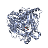
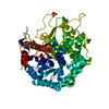
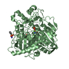
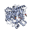
| Entry | Database: PDB / ID: 1hdh | |||||||||
|---|---|---|---|---|---|---|---|---|---|---|
| Title | Arylsulfatase from Pseudomonas aeruginosa | |||||||||
 Components Components | Arylsulfatase | |||||||||
 Keywords Keywords | HYDROLASE / SULFATASE / FORMYLGLYCINE HYDRATE | |||||||||
| Function / homology |  Function and homology information Function and homology informationarylsulfatase (type I) / arylsulfatase activity / phosphoric diester hydrolase activity / metal ion binding / cytoplasm Similarity search - Function | |||||||||
| Biological species |   Pseudomonas aeruginosa (bacteria) Pseudomonas aeruginosa (bacteria) | |||||||||
| Method |  X-RAY DIFFRACTION / SYNCHROTRON / SIRAS / Resolution: 1.3 Å X-RAY DIFFRACTION / SYNCHROTRON / SIRAS / Resolution: 1.3 Å | |||||||||
 Authors Authors | Boltes, I. / Czapinska, H. / Kahnert, A. / von Buelow, R. / Dirks, T. / Schmidt, B. / von Figura, K. / Kertesz, M.A. / Uson, I. | |||||||||
 Citation Citation | Journal: Structure / Year: 2001 Title: 1.3 A Structure of Arylsulfatase from Pseudomonas Aeruginosa Establishes the Catalytic Mechanism of Sulfate Ester Cleavage in the Sulfatase Family. Authors: Boltes, I. / Czapinska, H. / Kahnert, A. / von Buelow, R. / Dirks, T. / Schmidt, B. / von Figura, K. / Kertesz, M.A. / Uson, I. | |||||||||
| History |
|
- Structure visualization
| Structure viewer | Molecule: MolmilJmol/JSmol |
|---|
- Downloads & links
-Download
| PDBx/mmCIF format | 1hdh.cif.gz | 231.2 KB | Display | PDBx/mmCIF format |
|---|---|---|---|---|
| PDB format | pdb1hdh.ent.gz | 182.6 KB | Display | PDB format |
| PDBx/mmJSON format | 1hdh.json.gz | Tree view | PDBx/mmJSON format | |
| Others |  Other downloads Other downloads |
-Validation report
| Arichive directory | https://data.pdbj.org/pub/pdb/validation_reports/hd/1hdhftp://data.pdbj.org/pub/pdb/validation_reports/hd/1hdh | HTTPS FTP |
|---|
-Related structure data
| Similar structure data |
|---|


-Links
 PDBj
PDBj
- Assembly
| Deposited unit | 
| ||||||||
|---|---|---|---|---|---|---|---|---|---|
| 1 | 
| ||||||||
| 2 | 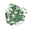
| ||||||||
| Unit cell |
| ||||||||
| Noncrystallographic symmetry (NCS) | NCS oper: (Code: given Matrix: (-0.98902, -0.00177, -0.14774), Vector: |
-Components
| #1: Protein | Mass: 60019.539 Da / Num. of mol.: 2 Source method: isolated from a genetically manipulated source Source: (gene. exp.) Pseudomonas aeruginosa (bacteria) / Gene: atsA, PA0183 / Production host: #2: Chemical |   Mass: 40.078 Da / Num. of mol.: 2 / Source method: obtained synthetically / Formula: Ca Mass: 40.078 Da / Num. of mol.: 2 / Source method: obtained synthetically / Formula: Ca#3: Chemical | ChemComp-SO4 /   Mass: 96.063 Da / Num. of mol.: 8 / Source method: obtained synthetically / Formula: SO4 Mass: 96.063 Da / Num. of mol.: 8 / Source method: obtained synthetically / Formula: SO4#4: Water | ChemComp-HOH / |  Mass: 18.015 Da / Num. of mol.: 810 / Source method: isolated from a natural source / Formula: H2O Mass: 18.015 Da / Num. of mol.: 810 / Source method: isolated from a natural source / Formula: H2OHas protein modification | Y | |
|---|
-Experimental details
-Experiment
| Experiment | Method: X-RAY DIFFRACTION / Number of used crystals: 1 |
|---|
- Sample preparation
| Crystal | Density Matthews: 2.1 Å3/Da / Density % sol: 40 % | ||||||||||||||||||||||||||||||||||||
|---|---|---|---|---|---|---|---|---|---|---|---|---|---|---|---|---|---|---|---|---|---|---|---|---|---|---|---|---|---|---|---|---|---|---|---|---|---|
| Crystal grow | Temperature: 293 K / Method: vapor diffusion, hanging drop / pH: 6.3 Details: PROTEIN SOLUTION: 11MG/ML IN 20 MM TRIS-HCL PH 7.5 + EQUAL VOLUME OF PRECIPITANT: 100 MM MES PH 6.3, 200 MM AMMONIUM SULFATE, 20% (W/V) PEG MONOMETHYLETHER 5000; HANGING DROP, 20 C | ||||||||||||||||||||||||||||||||||||
| Crystal grow | *PLUS Temperature: 18-22 ℃ / Method: vapor diffusion, hanging drop | ||||||||||||||||||||||||||||||||||||
| Components of the solutions | *PLUS
|
-Data collection
| Diffraction | Mean temperature: 113 K |
|---|---|
| Diffraction source | Source: SYNCHROTRON / Site: EMBL/DESY, HAMBURG  / Beamline: X11 / Wavelength: 0.91 / Beamline: X11 / Wavelength: 0.91 |
| Detector | Type: MARRESEARCH / Detector: IMAGE PLATE / Date: Aug 15, 1999 / Details: BENT CRYSTAL |
| Radiation | Monochromator: GE SINGLE CRYSTAL / Protocol: SINGLE WAVELENGTH / Monochromatic (M) / Laue (L): M / Scattering type: x-ray |
| Radiation wavelength | Wavelength: 0.91 Å / Relative weight: 1 |
| Reflection | Resolution: 1.3→20 Å / Num. obs: 255817 / % possible obs: 93.1 % / Redundancy: 1.7 % / Rmerge(I) obs: 0.062 / Net I/σ(I): 12.7 |
| Reflection shell | Resolution: 1.3→1.4 Å / Redundancy: 1.5 % / Rmerge(I) obs: 0.259 / Mean I/σ(I) obs: 3.5 / % possible all: 81.5 |
| Reflection | *PLUS Lowest resolution: 20 Å / Rmerge(I) obs: 0.085 |
| Reflection shell | *PLUS % possible obs: 81.5 % / Rmerge(I) obs: 0.337 |
- Processing
| Software |
| |||||||||||||||||||||||||||||||||
|---|---|---|---|---|---|---|---|---|---|---|---|---|---|---|---|---|---|---|---|---|---|---|---|---|---|---|---|---|---|---|---|---|---|---|
| Refinement | Method to determine structure: SIRAS / Resolution: 1.3→20 Å / Num. parameters: 36198 / Num. restraintsaints: 46182 / Cross valid method: FREE R-VALUE / σ(F): 0 / StereochEM target val spec case: CSD FOR DDZ 51 / Stereochemistry target values: ENGH AND HUBER
| |||||||||||||||||||||||||||||||||
| Solvent computation | Solvent model: MOEWS & KRETSINGER, J.MOL.BIOL.91(1973)201-228 | |||||||||||||||||||||||||||||||||
| Refine analyze | Num. disordered residues: 17 / Occupancy sum hydrogen: 7763.36 / Occupancy sum non hydrogen: 8989.8 | |||||||||||||||||||||||||||||||||
| Refinement step | Cycle: LAST / Resolution: 1.3→20 Å
| |||||||||||||||||||||||||||||||||
| Refine LS restraints |
| |||||||||||||||||||||||||||||||||
| Software | *PLUS Name: SHELXL-97 / Classification: refinement | |||||||||||||||||||||||||||||||||
| Refinement | *PLUS Rfactor obs: 0.1894 / Rfactor Rfree: 0.2179 / Rfactor Rwork: 0.1885 | |||||||||||||||||||||||||||||||||
| Solvent computation | *PLUS | |||||||||||||||||||||||||||||||||
| Displacement parameters | *PLUS |
