 Movie
Movie Controller


+ Open data

- Basic information
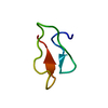
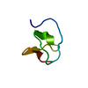
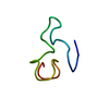
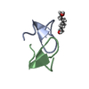
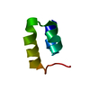
| Entry | Database: PDB / ID: 1hd6 | ||||||
|---|---|---|---|---|---|---|---|
| Title | PHEROMONE ER-22, NMR | ||||||
 Components Components | PHEROMONE ER-22 | ||||||
 Keywords Keywords | PHEROMONE | ||||||
| Function / homology | Protozoan pheromone superfamily / Pheromone, protozoan / Euplotes raikovi mating pheromone / pheromone activity / extracellular region / Mating pheromone Er-22 Function and homology information Function and homology information | ||||||
| Biological species |  EUPLOTES RAIKOVI (eukaryote) EUPLOTES RAIKOVI (eukaryote) | ||||||
| Method | SOLUTION NMR / DISTANCE GEOMETRY, RESTRAINED ENERGY REFINEMENT | ||||||
 Authors Authors | Luginbuhl, P. / Liu, A. / Zerbe, O. / Ortenzi, C. / Luporini, P. / Wuthrich, K. | ||||||
 Citation Citation | Journal: J.Biomol.NMR / Year: 2001 Title: NMR Structure of the Pheromone Er-22 from Euplotes Raikovi Authors: Liu, A. / Luginbuhl, P. / Zerbe, O. / Ortenzi, C. / Luporini, P. / Wuthrich, K. | ||||||
| History |
| ||||||
| Remark 650 | HELIX DETERMINATION METHOD: AUTHOR PROVIDED. |
- Structure visualization
| Structure viewer | Molecule: MolmilJmol/JSmol |
|---|
- Downloads & links
-Download
| PDBx/mmCIF format | 1hd6.cif.gz | 208.2 KB | Display | PDBx/mmCIF format |
|---|---|---|---|---|
| PDB format | pdb1hd6.ent.gz | 173.2 KB | Display | PDB format |
| PDBx/mmJSON format | 1hd6.json.gz | Tree view | PDBx/mmJSON format | |
| Others |  Other downloads Other downloads |
-Validation report
| Arichive directory | https://data.pdbj.org/pub/pdb/validation_reports/hd/1hd6ftp://data.pdbj.org/pub/pdb/validation_reports/hd/1hd6 | HTTPS FTP |
|---|
-Related structure data
| Similar structure data |
|---|


-Links
 PDBj
PDBj- Assembly
| Deposited unit | 
| |||||||||
|---|---|---|---|---|---|---|---|---|---|---|
| 1 |
| |||||||||
| NMR ensembles |
|
-Components
| #1: Protein/peptide | Mass: 3939.533 Da / Num. of mol.: 1 / Source method: isolated from a natural source / Source: (natural) EUPLOTES RAIKOVI (eukaryote) / References: UniProt: P58548*PLUS |
|---|---|
| Has protein modification | Y |
-Experimental details
-Experiment
| Experiment | Method: SOLUTION NMR | ||||||||||||||||||||||||||||
|---|---|---|---|---|---|---|---|---|---|---|---|---|---|---|---|---|---|---|---|---|---|---|---|---|---|---|---|---|---|
| NMR experiment |
| ||||||||||||||||||||||||||||
| NMR details | Text: REPRESENTATIVE CONFORMER HAS THE SMALLEST RMSD TO THE MEAN STRUCTURE UPON SUPERPOSITION OF THE BACKBONE ATOMS N, CA, AND C' OF RESIDUES 2-32. |
 HSQC
HSQC- Sample preparation
| Sample conditions | pH: 5 / Pressure: 1 atm / Temperature: 296 K |
|---|---|
| Crystal grow | *PLUS Method: other / Details: NMR |
-NMR measurement
| NMR spectrometer |
|
|---|
- Processing
| NMR software |
| ||||||||||||
|---|---|---|---|---|---|---|---|---|---|---|---|---|---|
| Refinement | Method: DISTANCE GEOMETRY, RESTRAINED ENERGY REFINEMENT / Software ordinal: 1 Details: REFINEMENT DETAILS CAN BE FOUND IN THE JRNL CITATION ABOVE. THREE-DIMENSIONAL STRUCTURE IN AQUEOUS SOLUTION REPRESENTED BY 20 CONFORMERS DETERMINED BY NUCLEAR MAGNETIC RESONANCE, DISTANCE ...Details: REFINEMENT DETAILS CAN BE FOUND IN THE JRNL CITATION ABOVE. THREE-DIMENSIONAL STRUCTURE IN AQUEOUS SOLUTION REPRESENTED BY 20 CONFORMERS DETERMINED BY NUCLEAR MAGNETIC RESONANCE, DISTANCE GEOMETRY AND RESTRAINED ENERGY REFINEMENT. DATA WERE COLLECTED AT 23 DEGREES CELSIUS AND AT PH 5.0. THEY CONSIST OF 497 UPPER LIMITS ON DISTANCES OBTAINED FROM NOE MEASUREMENTS AND 41 ANGLE CONSTRAINTS OBTAINED FROM NOE MEASUREMENTS AND COUPLING CONSTANT MEASUREMENTS. THESE INPUT DATA ARE ALSO AVAILABLE FROM THE PROTEIN DATA BANK. DISTANCE GEOMETRY CALCULATIONS WERE PERFORMED WITH THE PROGRAM DIANA (P. GUENTERT, W. BRAUN, K. WUTHRICH, J. MOL. BIOL. (1991) VOL. 217, 517 - 530). FOR THE RESTRAINED ENERGY MINIMIZATION THE PROGRAM OPAL (P. LUGINBUHL ET AL., J. BIOMOL. NMR (1996) VOL. 8, 136-146) WAS USED. FOR THE PRESENT STRUCTURES THE NMR DISTANCE CONSTRAINTS WERE WEIGHTED SUCH THAT A VIOLATION OF AN UPPER DISTANCE LIMIT OF 0.1 ANGSTROM CORRESPONDS TO AN ENERGY OF KT/2. THE CONSTRAINTS ON DIHEDRAL ANGLES RESULTING FROM MEASUREMENTS OF VICINAL COUPLING CONSTANTS WERE WEIGHTED SUCH THAT A VIOLATION OF 2.5 DEGREES CORRESPONDS TO AN ENERGY OF KT/2. DEPOSITED COORDINATES ARE THOSE OF CONFORMERS 1 - 20 IN THE PAPER CITED ON *JRNL* RECORDS ABOVE. NO VIOLATIONS OF DISTANCE CONSTRAINTS FROM NOES EXCEED 0.10 ANGSTROMS, AND NO VIOLATIONS OF ANGLE CONSTRAINTS EXCEED 2.5 DEGREES. ATOM NAMES HAVE BEEN ASSIGNED FOLLOWING THE RECOMMENDATIONS OF THE IUPAC-IUB COMMISSION AS PUBLISHED IN BIOCHEMISTRY (1970) VOL. 8, 3471 - 3479. THE INDIVIDUAL NUMBERS OF THE HYDROGEN ATOMS IN METHYL AND METHYLENE GROUPS ARE INDICATED AS THE FIRST CHARACTER RATHER THAN THE LAST CHARACTER OF THE ATOM NAMES. THE AVERAGE OF THE RMSD VALUES IN A PAIRWISE COMPARISON OF THE 20 NMR CONFORMERS TO THE MEAN STRUCTURE AS DESCRIBED IN THE PAPER CITED ON * JRNL* RECORDS ABOVE IS 0.47 ANGSTROMS FOR THE BACKBONE ATOMS OF RESIDUES 1 - 37, AND 0.75 ANGSTROMS FOR ALL HEAVY ATOMS. EXCLUDING THE CARBOXY-TERMINUS AND THE AMINO-TERMINUS, WHICH ARE LESS WELL DEFINED BY THE NMR DATA, THE AVERAGE OF THE RMSD VALUES IN A PAIRWISE COMPARISON TO THE MEAN STRUCTURE FOR RESIDUES 2 - 32 IS 0.27 ANGSTROMS FOR THE BACKBONE ATOMS. | ||||||||||||
| NMR ensemble | Conformer selection criteria: LOWEST RESIDUAL TARGET FUNCTION Conformers calculated total number: 50 / Conformers submitted total number: 20 |
