 Movie
Movie Controller

[English] 日本語
 Yorodumi
- PDB-1hcv: LLAMA HEAVY CHAIN VARIABLE DOMAIN AGAINST ALPHA SUBUNIT OF HCG (H... -
+ Open data

- Basic information
| Entry | Database: PDB / ID: 1hcv | ||||||
|---|---|---|---|---|---|---|---|
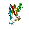
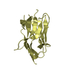
| Title | LLAMA HEAVY CHAIN VARIABLE DOMAIN AGAINST ALPHA SUBUNIT OF HCG (HUMAN CHORIONIC GONADOTROPIN) | ||||||
 Components Components | IMMUNOGLOBULIN G | ||||||
 Keywords Keywords | IMMUNOGLOBULIN / CAMELIDS | ||||||
| Function / homology | Immunoglobulins / Immunoglobulin-like / Sandwich / Mainly Beta / :  Function and homology information Function and homology information | ||||||
| Biological species |  | ||||||
| Method |  X-RAY DIFFRACTION / MOLECULAR REPLACEMENT / Resolution: 1.85 Å X-RAY DIFFRACTION / MOLECULAR REPLACEMENT / Resolution: 1.85 Å | ||||||
 Authors Authors | Spinelli, S. / Cambillau, C. / Tegoni, M. | ||||||
 Citation Citation | Journal: Nat.Struct.Biol. / Year: 1996 Title: The crystal structure of a llama heavy chain variable domain. Authors: Spinelli, S. / Frenken, L. / Bourgeois, D. / de Ron, L. / Bos, W. / Verrips, T. / Anguille, C. / Cambillau, C. / Tegoni, M. #1: Journal: Nat.Struct.Biol. / Year: 1996Title: Crystal Structure of a Camel Single-Domain Vh Antibody Fragment in Complex with Lysozyme Authors: Desmyter, A. / Transue, T.R. / Ghahroudi, M.A. / Thi, M.H. / Poortmans, F. / Hamers, R. / Muyldermans, S. / Wyns, L. #2: Journal: Nat.Struct.Biol. / Year: 1996Title: Redefining the Minimal Antigen-Binding Fragment Authors: Sheriff, S. / Constantine, K.L. #3: Journal: Protein Eng. / Year: 1994Title: Sequence and Structure of Vh Domain from Naturally Occurring Camel Heavy Chain Immunoglobulins Lacking Light Chains Authors: Muyldermans, S. / Atarhouch, T. / Saldanha, J. / Barbosa, J.A. / Hamers, R. #4: Journal: Nature / Year: 1993Title: Naturally Occurring Antibodies Devoid of Light Chains Authors: Hamers-Casterman, C. / Atarhouch, T. / Muyldermans, S. / Robinson, G. / Hamers, C. / Songa, E.B. / Bendahman, N. / Hamers, R. | ||||||
| History |
|
- Structure visualization
| Structure viewer | Molecule: MolmilJmol/JSmol |
|---|
- Downloads & links
-Download
| PDBx/mmCIF format | 1hcv.cif.gz | 44.1 KB | Display | PDBx/mmCIF format |
|---|---|---|---|---|
| PDB format | pdb1hcv.ent.gz | 31.2 KB | Display | PDB format |
| PDBx/mmJSON format | 1hcv.json.gz | Tree view | PDBx/mmJSON format | |
| Others |  Other downloads Other downloads |
-Validation report
| Summary document | 1hcv_validation.pdf.gz | 359.2 KB | Display | wwPDB validaton report |
|---|---|---|---|---|
| Full document | 1hcv_full_validation.pdf.gz | 362 KB | Display | |
| Data in XML | 1hcv_validation.xml.gz | 3.8 KB | Display | |
| Data in CIF | 1hcv_validation.cif.gz | 5.8 KB | Display | |
| Arichive directory | https://data.pdbj.org/pub/pdb/validation_reports/hc/1hcvftp://data.pdbj.org/pub/pdb/validation_reports/hc/1hcv | HTTPS FTP |
-Related structure data
| Similar structure data |
|---|


-Links
 PDBj
PDBj

- Assembly
| Deposited unit | 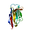
| ||||||||
|---|---|---|---|---|---|---|---|---|---|
| 1 |
| ||||||||
| Unit cell |
|
-Components
| #1: Antibody | Mass: 12508.674 Da / Num. of mol.: 1 / Fragment: HEAVY CHAIN VARIABLE DOMAIN FROM LLAMA Source method: isolated from a genetically manipulated source Details: FROM IMMUNIZED LLAMA / Source: (gene. exp.)  |
|---|---|
| #2: Water | ChemComp-HOH /  Mass: 18.015 Da / Num. of mol.: 103 / Source method: isolated from a natural source / Formula: H2O Mass: 18.015 Da / Num. of mol.: 103 / Source method: isolated from a natural source / Formula: H2O |
-Experimental details
-Experiment
| Experiment | Method: X-RAY DIFFRACTION / Number of used crystals: 2 |
|---|
- Sample preparation
| Crystal | Density Matthews: 1.72 Å3/Da / Density % sol: 32 % | ||||||||||||||||||||
|---|---|---|---|---|---|---|---|---|---|---|---|---|---|---|---|---|---|---|---|---|---|
| Crystal grow | Method: vapor diffusion / pH: 8 Details: VAPOR DIFFUSION, PEG 8K 25%, TRIS 0.1M, PH 8.0, vapor diffusion | ||||||||||||||||||||
| Crystal grow | *PLUS Temperature: 20 ℃ / Method: vapor diffusion | ||||||||||||||||||||
| Components of the solutions | *PLUS
|
-Data collection
| Diffraction | Mean temperature: 290 K |
|---|---|
| Diffraction source | Source: ROTATING ANODE / Type: RIGAKU FR-D / Wavelength: 1.5418 |
| Detector | Type: MARRESEARCH / Detector: IMAGE PLATE / Date: Feb 19, 1996 |
| Radiation | Monochromator: GRAPHITE(002) / Monochromatic (M) / Laue (L): M / Scattering type: x-ray |
| Radiation wavelength | Wavelength: 1.5418 Å / Relative weight: 1 |
| Reflection | Resolution: 2.5→30 Å / Num. obs: 4270 / % possible obs: 98 % / Observed criterion σ(I): 0 / Redundancy: 1.6 % / Rmerge(I) obs: 0.109 / Net I/σ(I): 9.03 |
| Reflection shell | Resolution: 2.5→2.59 Å / Redundancy: 1.9 % / Rmerge(I) obs: 0.17 / Mean I/σ(I) obs: 3.4 / % possible all: 98.7 |
| Reflection | *PLUS Num. obs: 8077 / % possible obs: 99.8 % / Num. measured all: 92439 / Rmerge(I) obs: 0.089 |
| Reflection shell | *PLUS Highest resolution: 1.85 Å / Lowest resolution: 1.88 Å / % possible obs: 100 % / Rmerge(I) obs: 0.191 / Mean I/σ(I) obs: 10.4 |
- Processing
| Software |
| ||||||||||||||||||||||||||||||||||||||||||||||||||||||||||||
|---|---|---|---|---|---|---|---|---|---|---|---|---|---|---|---|---|---|---|---|---|---|---|---|---|---|---|---|---|---|---|---|---|---|---|---|---|---|---|---|---|---|---|---|---|---|---|---|---|---|---|---|---|---|---|---|---|---|---|---|---|---|
| Refinement | Method to determine structure: MOLECULAR REPLACEMENT Starting model: VH OF FAB FRAGMENT OF ANTIBODY AGAINST 2-PHENYL-OXAZOLONE Resolution: 1.85→6 Å / Cross valid method: YES / σ(F): 1
| ||||||||||||||||||||||||||||||||||||||||||||||||||||||||||||
| Displacement parameters | Biso mean: 16 Å2 | ||||||||||||||||||||||||||||||||||||||||||||||||||||||||||||
| Refinement step | Cycle: LAST / Resolution: 1.85→6 Å
| ||||||||||||||||||||||||||||||||||||||||||||||||||||||||||||
| Refine LS restraints |
| ||||||||||||||||||||||||||||||||||||||||||||||||||||||||||||
| Software | *PLUS Name: X-PLOR / Classification: refinement | ||||||||||||||||||||||||||||||||||||||||||||||||||||||||||||
| Refinement | *PLUS | ||||||||||||||||||||||||||||||||||||||||||||||||||||||||||||
| Solvent computation | *PLUS | ||||||||||||||||||||||||||||||||||||||||||||||||||||||||||||
| Displacement parameters | *PLUS | ||||||||||||||||||||||||||||||||||||||||||||||||||||||||||||
| Refine LS restraints | *PLUS
|
