 Movie
Movie Controller

[English] 日本語
 Yorodumi
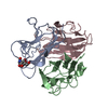
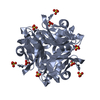
- PDB-1h9m: Two crystal structures of the cytoplasmic molybdate-binding prote... -
+ Open data

- Basic information
| Entry | Database: PDB / ID: 1h9m | ||||||
|---|---|---|---|---|---|---|---|
| Title | Two crystal structures of the cytoplasmic molybdate-binding protein ModG suggest a novel cooperative binding mechanism and provide insights into ligand-binding specificity. PEG-grown form with molybdate bound | ||||||
 Components Components | MOLYBDENUM-BINDING-PROTEIN | ||||||
 Keywords Keywords | BINDING PROTEIN / MOLYBDATE HOMEOSTASIS | ||||||
| Function / homology |  Function and homology information Function and homology information | ||||||
| Biological species |  AZOTOBACTER VINELANDII (bacteria) AZOTOBACTER VINELANDII (bacteria) | ||||||
| Method |  X-RAY DIFFRACTION / SYNCHROTRON / MOLECULAR REPLACEMENT / Resolution: 1.65 Å X-RAY DIFFRACTION / SYNCHROTRON / MOLECULAR REPLACEMENT / Resolution: 1.65 Å | ||||||
 Authors Authors | Delarbre, L. / Stevenson, C.E.M. / White, D.J. / Mitchenall, L.A. / Pau, R.N. / Lawson, D.M. | ||||||
 Citation Citation | Journal: J.Mol.Biol. / Year: 2001 Title: Two Crystal Structures of the Cytoplasmic Molybdate-Binding Protein Modg Suggest a Novel Cooperative Binding Mechanism and Provide Insights Into Ligand-Binding Specificity Authors: Delarbre, L. / Stevenson, C.E.M. / White, D.J. / Mitchenall, L.A. / Pau, R.N. / Lawson, D.M. | ||||||
| History |
|
- Structure visualization
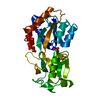
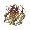
| Structure viewer | Molecule: MolmilJmol/JSmol |
|---|
- Downloads & links
-Download
| PDBx/mmCIF format | 1h9m.cif.gz | 66.3 KB | Display | PDBx/mmCIF format |
|---|---|---|---|---|
| PDB format | pdb1h9m.ent.gz | 49 KB | Display | PDB format |
| PDBx/mmJSON format | 1h9m.json.gz | Tree view | PDBx/mmJSON format | |
| Others |  Other downloads Other downloads |
-Validation report
| Arichive directory | https://data.pdbj.org/pub/pdb/validation_reports/h9/1h9mftp://data.pdbj.org/pub/pdb/validation_reports/h9/1h9m | HTTPS FTP |
|---|
-Related structure data
| Related structure data | 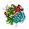 1h9jSC  1h9kC S: Starting model for refinement C: citing same article ( |
|---|---|
| Similar structure data |


-Links
 PDBj
PDBj- Assembly
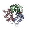
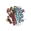
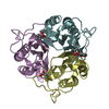
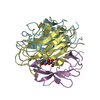
| Deposited unit | 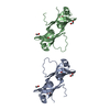
| |||||||||||||||||||||||||||
|---|---|---|---|---|---|---|---|---|---|---|---|---|---|---|---|---|---|---|---|---|---|---|---|---|---|---|---|---|
| 1 | 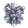
| |||||||||||||||||||||||||||
| 2 | 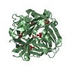
| |||||||||||||||||||||||||||
| Unit cell |
| |||||||||||||||||||||||||||
| Components on special symmetry positions |
| |||||||||||||||||||||||||||
| Noncrystallographic symmetry (NCS) | NCS oper: (Code: given Matrix: (-0.50232, 0.86468, -0.00062), Vector: Details | BIOMOLECULE | |
-Components
| #1: Protein | Mass: 14654.964 Da / Num. of mol.: 2 Source method: isolated from a genetically manipulated source Source: (gene. exp.) AZOTOBACTER VINELANDII (bacteria) / Strain: E162 / Cellular location: CYTOPLASM / Gene: MODG / Gene (production host): MODG / Production host: #2: Chemical | ChemComp-MOO /   Mass: 159.938 Da / Num. of mol.: 8 / Source method: obtained synthetically / Formula: MoO4 Mass: 159.938 Da / Num. of mol.: 8 / Source method: obtained synthetically / Formula: MoO4#3: Water | ChemComp-HOH / |  Mass: 18.015 Da / Num. of mol.: 79 / Source method: isolated from a natural source / Formula: H2O Mass: 18.015 Da / Num. of mol.: 79 / Source method: isolated from a natural source / Formula: H2O |
|---|
-Experimental details
-Experiment
| Experiment | Method: X-RAY DIFFRACTION / Number of used crystals: 1 |
|---|
- Sample preparation
| Crystal | Density Matthews: 1.34 Å3/Da / Density % sol: 41 % | |||||||||||||||||||||||||||||||||||
|---|---|---|---|---|---|---|---|---|---|---|---|---|---|---|---|---|---|---|---|---|---|---|---|---|---|---|---|---|---|---|---|---|---|---|---|---|
| Crystal grow | Method: vapor diffusion / pH: 7.5 Details: VAPOUR DIFFUSION. 20% PEG 4000, 5% ISOPROPANOL IN 100MM HEPES PH7.5 WITH 2MM NA2MOO4., pH 7.50 | |||||||||||||||||||||||||||||||||||
| Crystal grow | *PLUS Method: vapor diffusion / pH: 7.5 | |||||||||||||||||||||||||||||||||||
| Components of the solutions | *PLUS
|
-Data collection
| Diffraction | Mean temperature: 100 K |
|---|---|
| Diffraction source | Source: SYNCHROTRON / Site: ESRF  / Beamline: ID14-2 / Wavelength: 0.933 / Beamline: ID14-2 / Wavelength: 0.933 |
| Detector | Type: MARRESEARCH / Detector: CCD / Date: Jun 15, 1999 |
| Radiation | Protocol: SINGLE WAVELENGTH / Monochromatic (M) / Laue (L): M / Scattering type: x-ray |
| Radiation wavelength | Wavelength: 0.933 Å / Relative weight: 1 |
| Reflection | Resolution: 1.65→41 Å / Num. obs: 28231 / % possible obs: 100 % / Observed criterion σ(I): -3 / Redundancy: 12.4 % / Biso Wilson estimate: 19.9 Å2 / Rmerge(I) obs: 0.044 / Net I/σ(I): 41.7 |
| Reflection shell | Resolution: 1.65→1.68 Å / Rmerge(I) obs: 0.107 / Mean I/σ(I) obs: 8.8 / % possible all: 100 |
| Reflection shell | *PLUS % possible obs: 100 % |
- Processing
| Software |
| ||||||||||||||||||||||||||||||||||||||||||||||||||||||||||||||||||||||||||||||||||||
|---|---|---|---|---|---|---|---|---|---|---|---|---|---|---|---|---|---|---|---|---|---|---|---|---|---|---|---|---|---|---|---|---|---|---|---|---|---|---|---|---|---|---|---|---|---|---|---|---|---|---|---|---|---|---|---|---|---|---|---|---|---|---|---|---|---|---|---|---|---|---|---|---|---|---|---|---|---|---|---|---|---|---|---|---|---|
| Refinement | Method to determine structure: MOLECULAR REPLACEMENT Starting model: 1H9J Resolution: 1.65→40 Å / Cross valid method: THROUGHOUT / σ(F): 0 / ESU R: 0.111 / ESU R Free: 0.108
| ||||||||||||||||||||||||||||||||||||||||||||||||||||||||||||||||||||||||||||||||||||
| Displacement parameters | Biso mean: 27 Å2 | ||||||||||||||||||||||||||||||||||||||||||||||||||||||||||||||||||||||||||||||||||||
| Refinement step | Cycle: LAST / Resolution: 1.65→40 Å
| ||||||||||||||||||||||||||||||||||||||||||||||||||||||||||||||||||||||||||||||||||||
| Refine LS restraints |
| ||||||||||||||||||||||||||||||||||||||||||||||||||||||||||||||||||||||||||||||||||||
| Software | *PLUS Name: REFMAC / Classification: refinement | ||||||||||||||||||||||||||||||||||||||||||||||||||||||||||||||||||||||||||||||||||||
| Refinement | *PLUS Rfactor obs: 0.191 | ||||||||||||||||||||||||||||||||||||||||||||||||||||||||||||||||||||||||||||||||||||
| Solvent computation | *PLUS | ||||||||||||||||||||||||||||||||||||||||||||||||||||||||||||||||||||||||||||||||||||
| Displacement parameters | *PLUS |
