 Movie
Movie Controller


+ Open data

- Basic information
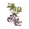
| Entry | Database: PDB / ID: 1h72 | |||||||||
|---|---|---|---|---|---|---|---|---|---|---|
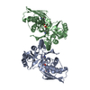
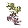
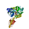
| Title | CRYSTAL STRUCTURE OF HOMOSERINE KINASE COMPLEXED WITH HSE | |||||||||
 Components Components | HOMOSERINE KINASE | |||||||||
 Keywords Keywords | TRANSFERASE / KINASE / THREONINE BIOSYNTHESIS | |||||||||
| Function / homology |  Function and homology information Function and homology informationhomoserine kinase / homoserine kinase activity / L-threonine biosynthetic process / ATP binding / cytoplasm Similarity search - Function | |||||||||
| Biological species |   METHANOCOCCUS JANNASCHII (archaea) METHANOCOCCUS JANNASCHII (archaea) | |||||||||
| Method |  X-RAY DIFFRACTION / SYNCHROTRON / MOLECULAR REPLACEMENT / Resolution: 1.8 Å X-RAY DIFFRACTION / SYNCHROTRON / MOLECULAR REPLACEMENT / Resolution: 1.8 Å | |||||||||
 Authors Authors | Krishna, S.S. / Zhou, T. / Daugherty, M. / Osterman, A.L. / Zhang, H. | |||||||||
 Citation Citation | Journal: Biochemistry / Year: 2001 Title: Structural Basis for the Catalysis and Substrate Specificity of Homoserine Kinase Authors: Krishna, S.S. / Zhou, T. / Daugherty, M. / Osterman, A.L. / Zhang, H. #1: Journal: Structure / Year: 2000Title: Structure and Mechanism of Homoserine Kinase: Prototype for the Ghmp Kinase Superfamily Authors: Zhou, T. / Daugherty, M. / Grishin, N.V. / Osterman, A.L. / Zhang, H. | |||||||||
| History |
|
- Structure visualization
| Structure viewer | Molecule: MolmilJmol/JSmol |
|---|
- Downloads & links
-Download
| PDBx/mmCIF format | 1h72.cif.gz | 78.3 KB | Display | PDBx/mmCIF format |
|---|---|---|---|---|
| PDB format | pdb1h72.ent.gz | 58.7 KB | Display | PDB format |
| PDBx/mmJSON format | 1h72.json.gz | Tree view | PDBx/mmJSON format | |
| Others |  Other downloads Other downloads |
-Validation report
| Arichive directory | https://data.pdbj.org/pub/pdb/validation_reports/h7/1h72ftp://data.pdbj.org/pub/pdb/validation_reports/h7/1h72 | HTTPS FTP |
|---|
-Related structure data
| Related structure data | 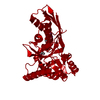 1h73C 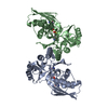 1h74C 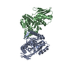 1fwlS S: Starting model for refinement C: citing same article ( |
|---|---|
| Similar structure data |


-Links
 PDBj
PDBj


- Assembly
| Deposited unit | 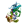
| ||||||||
|---|---|---|---|---|---|---|---|---|---|
| 1 | 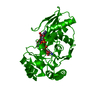
| ||||||||
| Unit cell |
| ||||||||
| Details | THE CHAIN IN THE COMPLEX IS 1422.9TROM **2 |
-Components
| #1: Protein | Mass: 32299.371 Da / Num. of mol.: 1 Source method: isolated from a genetically manipulated source Source: (gene. exp.) METHANOCOCCUS JANNASCHII (archaea) / Production host:  |
|---|---|
| #2: Chemical | ChemComp-HSE /   Type: L-peptide linking / Mass: 119.119 Da / Num. of mol.: 1 / Source method: obtained synthetically / Formula: C4H9NO3 Type: L-peptide linking / Mass: 119.119 Da / Num. of mol.: 1 / Source method: obtained synthetically / Formula: C4H9NO3 |
| #3: Chemical | ChemComp-ANP /   Mass: 506.196 Da / Num. of mol.: 1 / Source method: obtained synthetically / Formula: C10H17N6O12P3 / Comment: AMP-PNP, energy-carrying molecule analogue*YM Mass: 506.196 Da / Num. of mol.: 1 / Source method: obtained synthetically / Formula: C10H17N6O12P3 / Comment: AMP-PNP, energy-carrying molecule analogue*YM |
| #4: Chemical | ChemComp-TRS /   Mass: 122.143 Da / Num. of mol.: 1 / Source method: obtained synthetically / Formula: C4H12NO3 / Comment: pH buffer*YM Mass: 122.143 Da / Num. of mol.: 1 / Source method: obtained synthetically / Formula: C4H12NO3 / Comment: pH buffer*YM |
| #5: Water | ChemComp-HOH /  Mass: 18.015 Da / Num. of mol.: 308 / Source method: isolated from a natural source / Formula: H2O Mass: 18.015 Da / Num. of mol.: 308 / Source method: isolated from a natural source / Formula: H2O |
| Has protein modification | N |
-Experimental details
-Experiment
| Experiment | Method: X-RAY DIFFRACTION / Number of used crystals: 1 |
|---|
- Sample preparation
| Crystal | Density Matthews: 2.98 Å3/Da / Density % sol: 58.66 % | ||||||||||||||||||||||||||||||||||||||||||||||||||||||||||||
|---|---|---|---|---|---|---|---|---|---|---|---|---|---|---|---|---|---|---|---|---|---|---|---|---|---|---|---|---|---|---|---|---|---|---|---|---|---|---|---|---|---|---|---|---|---|---|---|---|---|---|---|---|---|---|---|---|---|---|---|---|---|
| Crystal grow | pH: 8 / Details: PEG4000, SODIUM ACETATE, pH 8.00 | ||||||||||||||||||||||||||||||||||||||||||||||||||||||||||||
| Crystal grow | *PLUS Temperature: 100 K / pH: 7.2 / Method: vapor diffusion, hanging drop | ||||||||||||||||||||||||||||||||||||||||||||||||||||||||||||
| Components of the solutions | *PLUS
|
-Data collection
| Diffraction | Mean temperature: 100 K |
|---|---|
| Diffraction source | Source: SYNCHROTRON / Site: APS  / Beamline: 19-BM / Wavelength: 0.9793 / Beamline: 19-BM / Wavelength: 0.9793 |
| Detector | Detector: IMAGE PLATE |
| Radiation | Protocol: SINGLE WAVELENGTH / Monochromatic (M) / Laue (L): M / Scattering type: x-ray |
| Radiation wavelength | Wavelength: 0.9793 Å / Relative weight: 1 |
| Reflection | Resolution: 1.8→50 Å / Num. obs: 36728 / % possible obs: 99.8 % / Observed criterion σ(I): -3 / Redundancy: 5.1 % / Rmerge(I) obs: 0.089 / Net I/σ(I): 21.1 |
| Reflection | *PLUS Num. measured all: 206493 |
| Reflection shell | *PLUS % possible obs: 99.9 % / Mean I/σ(I) obs: 2 |
- Processing
| Software |
| ||||||||||||||||||||||||||||||||||||||||||||||||||||||||||||
|---|---|---|---|---|---|---|---|---|---|---|---|---|---|---|---|---|---|---|---|---|---|---|---|---|---|---|---|---|---|---|---|---|---|---|---|---|---|---|---|---|---|---|---|---|---|---|---|---|---|---|---|---|---|---|---|---|---|---|---|---|---|
| Refinement | Method to determine structure: MOLECULAR REPLACEMENT Starting model: 1FWL Resolution: 1.8→50 Å / Cross valid method: FREE R-VALUE / σ(F): 0
| ||||||||||||||||||||||||||||||||||||||||||||||||||||||||||||
| Displacement parameters | Biso mean: 24.99 Å2 | ||||||||||||||||||||||||||||||||||||||||||||||||||||||||||||
| Refinement step | Cycle: LAST / Resolution: 1.8→50 Å
| ||||||||||||||||||||||||||||||||||||||||||||||||||||||||||||
| Refine LS restraints |
|
