 Movie
Movie Controller


+ Open data

- Basic information
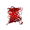
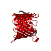
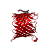
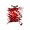
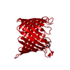
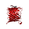
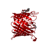
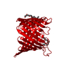
| Entry | Database: PDB / ID: 1h6s | ||||||
|---|---|---|---|---|---|---|---|
| Title | Asymmetric conductivity of engineered proteins | ||||||
 Components Components | PORIN | ||||||
 Keywords Keywords | MEMBRANE PROTEIN / INTEGRAL MEMBRANE PROTEIN PORIN | ||||||
| Function / homology |  Function and homology information Function and homology informationporin activity / pore complex / monoatomic ion transport / cell outer membrane Similarity search - Function | ||||||
| Biological species |  RHODOPSEUDOMONAS BLASTICA (bacteria) RHODOPSEUDOMONAS BLASTICA (bacteria) | ||||||
| Method |  X-RAY DIFFRACTION / MOLECULAR REPLACEMENT / Resolution: 3 Å X-RAY DIFFRACTION / MOLECULAR REPLACEMENT / Resolution: 3 Å | ||||||
 Authors Authors | Bannwarth, M. / Schulz, G.E. | ||||||
 Citation Citation | Journal: Protein Eng. / Year: 2002 Title: Asymmetric Conductivity of Engineered Porins Authors: Bannwarth, M. / Schulz, G.E. | ||||||
| History |
| ||||||
| Remark 700 | SHEET DETERMINATION METHOD: DSSP THE SHEETS PRESENTED AS "1A" IN EACH CHAIN ON SHEET RECORDS BELOW ... SHEET DETERMINATION METHOD: DSSP THE SHEETS PRESENTED AS "1A" IN EACH CHAIN ON SHEET RECORDS BELOW IS ACTUALLY AN 16-STRANDED BARREL THIS IS REPRESENTED BY A 17-STRANDED SHEET IN WHICH THE FIRST AND LAST STRANDS ARE IDENTICAL. |
- Structure visualization
| Structure viewer | Molecule: MolmilJmol/JSmol |
|---|
- Downloads & links
-Download
| PDBx/mmCIF format | 1h6s.cif.gz | 65.1 KB | Display | PDBx/mmCIF format |
|---|---|---|---|---|
| PDB format | pdb1h6s.ent.gz | 48 KB | Display | PDB format |
| PDBx/mmJSON format | 1h6s.json.gz | Tree view | PDBx/mmJSON format | |
| Others |  Other downloads Other downloads |
-Validation report
| Arichive directory | https://data.pdbj.org/pub/pdb/validation_reports/h6/1h6sftp://data.pdbj.org/pub/pdb/validation_reports/h6/1h6s | HTTPS FTP |
|---|
-Related structure data
| Related structure data |  1prnS S: Starting model for refinement |
|---|---|
| Similar structure data |


-Links
 PDBj
PDBj- Assembly
| Deposited unit | 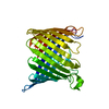
| ||||||||
|---|---|---|---|---|---|---|---|---|---|
| 1 | 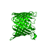
| ||||||||
| Unit cell |
|
-Components
| #1: Protein | Mass: 32222.725 Da / Num. of mol.: 1 / Mutation: YES Source method: isolated from a genetically manipulated source Details: INSERT SEQUENCE, GGGGPKLAKMEKARGGGG / Source: (gene. exp.) RHODOPSEUDOMONAS BLASTICA (bacteria)Description: RECOMBINANT ESCHERICHIA COLI. SYNTHETIC DNA INSERTION Plasmid: PET-3B-INS41L / Production host: |
|---|---|
| Compound details | CHAIN 1 AN 18 AMINO ACID INSERTION BETWEEN ASN 203 AND ASP 203 CHAIN 1 ENGINEERED MUTAION E1M: ...CHAIN 1 AN 18 AMINO ACID INSERTION BETWEEN ASN 203 AND ASP 203 CHAIN 1 ENGINEERED |
-Experimental details
-Experiment
| Experiment | Method: X-RAY DIFFRACTION / Number of used crystals: 1 |
|---|
- Sample preparation
| Crystal | Density Matthews: 4.05 Å3/Da / Density % sol: 69.63 % | |||||||||||||||||||||||||||||||||||||||||||||||||
|---|---|---|---|---|---|---|---|---|---|---|---|---|---|---|---|---|---|---|---|---|---|---|---|---|---|---|---|---|---|---|---|---|---|---|---|---|---|---|---|---|---|---|---|---|---|---|---|---|---|---|
| Crystal grow | pH: 4.8 Details: 50 MM NAOAC PH 4.75, 100 MM (NH4)2SO4, 5.5 % PEG4000 | |||||||||||||||||||||||||||||||||||||||||||||||||
| Crystal grow | *PLUS pH: 4.75 / Method: unknown | |||||||||||||||||||||||||||||||||||||||||||||||||
| Components of the solutions | *PLUS
|
-Data collection
| Diffraction | Mean temperature: 287 K |
|---|---|
| Diffraction source | Source: ROTATING ANODE / Type: RIGAKU RU200B / Wavelength: 1.5418 |
| Detector | Type: SIEMENS / Detector: IMAGE PLATE / Date: Oct 14, 2000 |
| Radiation | Monochromator: NI-FILTER / Protocol: SINGLE WAVELENGTH / Monochromatic (M) / Laue (L): M / Scattering type: x-ray |
| Radiation wavelength | Wavelength: 1.5418 Å / Relative weight: 1 |
| Reflection | Resolution: 3→36 Å / Num. obs: 10136 / % possible obs: 99 % / Observed criterion σ(I): 3 / Redundancy: 6.6 % / Biso Wilson estimate: 37.904 Å2 / Net I/σ(I): 19.1 |
| Reflection shell | Resolution: 3→3.03 Å / Redundancy: 6.5 % / Mean I/σ(I) obs: 5.1 / % possible all: 99 |
| Reflection | *PLUS % possible obs: 99 % / Num. measured all: 66659 / Rmerge(I) obs: 0.081 |
| Reflection shell | *PLUS Highest resolution: 3 Å / Lowest resolution: 3.15 Å / Num. unique obs: 1498 / Num. measured obs: 9696 / Rmerge(I) obs: 0.184 |
- Processing
| Software |
| ||||||||||||||||||||||||||||||||||||||||||||||||||||||||||||
|---|---|---|---|---|---|---|---|---|---|---|---|---|---|---|---|---|---|---|---|---|---|---|---|---|---|---|---|---|---|---|---|---|---|---|---|---|---|---|---|---|---|---|---|---|---|---|---|---|---|---|---|---|---|---|---|---|---|---|---|---|---|
| Refinement | Method to determine structure: MOLECULAR REPLACEMENT Starting model: PDB ENTRY 1PRN Resolution: 3→36 Å / Data cutoff high absF: 10000 / Cross valid method: RMSD / σ(F): 3 Details: INSERT REGION IS DISORDERED ONLY MISSING BOND IN COMPARISON TO WT IS SEEN.
| ||||||||||||||||||||||||||||||||||||||||||||||||||||||||||||
| Displacement parameters | Biso mean: 22.268 Å2 | ||||||||||||||||||||||||||||||||||||||||||||||||||||||||||||
| Refine analyze | Luzzati coordinate error obs: 0.2882 Å / Luzzati d res low obs: 5 Å / Luzzati sigma a obs: 0.2253 Å | ||||||||||||||||||||||||||||||||||||||||||||||||||||||||||||
| Refinement step | Cycle: LAST / Resolution: 3→36 Å
| ||||||||||||||||||||||||||||||||||||||||||||||||||||||||||||
| Refine LS restraints |
| ||||||||||||||||||||||||||||||||||||||||||||||||||||||||||||
| LS refinement shell | Resolution: 3→3.14 Å / Total num. of bins used: 8
| ||||||||||||||||||||||||||||||||||||||||||||||||||||||||||||
| Refinement | *PLUS Rfactor Rfree: 0.297 / Rfactor Rwork: 0.21 | ||||||||||||||||||||||||||||||||||||||||||||||||||||||||||||
| Solvent computation | *PLUS | ||||||||||||||||||||||||||||||||||||||||||||||||||||||||||||
| Displacement parameters | *PLUS | ||||||||||||||||||||||||||||||||||||||||||||||||||||||||||||
| Refine LS restraints | *PLUS
|
