 Movie
Movie Controller


+ Open data

- Basic information
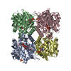
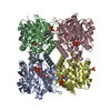
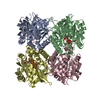
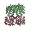
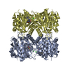
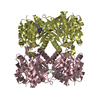
| Entry | Database: PDB / ID: 1h5s | ||||||
|---|---|---|---|---|---|---|---|
| Title | Thymidylyltransferase complexed with TMP | ||||||
 Components Components | (Glucose-1-phosphate thymidylyltransferase ...) x 4 | ||||||
 Keywords Keywords | TRANSFERASE / PYROPHOSPHATASE / NUCLEOTIDE SUGAR METHABOLISM | ||||||
| Function / homology |  Function and homology information Function and homology informationglucose-1-phosphate thymidylyltransferase / glucose-1-phosphate thymidylyltransferase activity / O antigen biosynthetic process / dTDP-rhamnose biosynthetic process / polysaccharide biosynthetic process / protein homotetramerization / metal ion binding / identical protein binding / cytosol Similarity search - Function | ||||||
| Biological species |  | ||||||
| Method |  X-RAY DIFFRACTION / SYNCHROTRON / MOLECULAR REPLACEMENT / Resolution: 2.3 Å X-RAY DIFFRACTION / SYNCHROTRON / MOLECULAR REPLACEMENT / Resolution: 2.3 Å | ||||||
 Authors Authors | Rosano, C. / Zuccotti, S. / Bolognesi, M. | ||||||
 Citation Citation | Journal: J. Mol. Biol. / Year: 2001 Title: Kinetic and crystallographic analyses support a sequential-ordered bi bi catalytic mechanism for Escherichia coli glucose-1-phosphate thymidylyltransferase. Authors: Zuccotti, S. / Zanardi, D. / Rosano, C. / Sturla, L. / Tonetti, M. / Bolognesi, M. | ||||||
| History |
|
- Structure visualization
| Structure viewer | Molecule: MolmilJmol/JSmol |
|---|
- Downloads & links
-Download
| PDBx/mmCIF format | 1h5s.cif.gz | 246.8 KB | Display | PDBx/mmCIF format |
|---|---|---|---|---|
| PDB format | pdb1h5s.ent.gz | 199 KB | Display | PDB format |
| PDBx/mmJSON format | 1h5s.json.gz | Tree view | PDBx/mmJSON format | |
| Others |  Other downloads Other downloads |
-Validation report
| Arichive directory | https://data.pdbj.org/pub/pdb/validation_reports/h5/1h5sftp://data.pdbj.org/pub/pdb/validation_reports/h5/1h5s | HTTPS FTP |
|---|
-Related structure data


-Links
 PDBj
PDBj

- Assembly
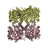
| Deposited unit | 
| ||||||||
|---|---|---|---|---|---|---|---|---|---|
| 1 |
| ||||||||
| Unit cell |
|
-Components
-Glucose-1-phosphate thymidylyltransferase ... , 4 types, 4 molecules ABCD
| #1: Protein | Mass: 32693.457 Da / Num. of mol.: 1 Source method: isolated from a genetically manipulated source Source: (gene. exp.) References: UniProt: P37744, glucose-1-phosphate thymidylyltransferase |
|---|---|
| #2: Protein | Mass: 32705.363 Da / Num. of mol.: 1 Source method: isolated from a genetically manipulated source Source: (gene. exp.) References: UniProt: P37744, glucose-1-phosphate thymidylyltransferase |
| #3: Protein | Mass: 32726.465 Da / Num. of mol.: 1 Source method: isolated from a genetically manipulated source Source: (gene. exp.) References: UniProt: P37744, glucose-1-phosphate thymidylyltransferase |
| #4: Protein | Mass: 32722.453 Da / Num. of mol.: 1 Source method: isolated from a genetically manipulated source Source: (gene. exp.) References: UniProt: P37744, glucose-1-phosphate thymidylyltransferase |
-Non-polymers , 2 types, 400 molecules 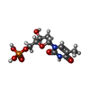

| #5: Chemical | ChemComp-TMP /  Mass: 322.208 Da / Num. of mol.: 8 / Source method: obtained synthetically / Formula: C10H15N2O8P Mass: 322.208 Da / Num. of mol.: 8 / Source method: obtained synthetically / Formula: C10H15N2O8P#6: Water | ChemComp-HOH / | Mass: 18.015 Da / Num. of mol.: 392 / Source method: isolated from a natural source / Formula: H2O |
|---|
-Experimental details
-Experiment
| Experiment | Method: X-RAY DIFFRACTION / Number of used crystals: 1 |
|---|
- Sample preparation
| Crystal | Density Matthews: 2.48 Å3/Da / Density % sol: 50.4 % |
|---|---|
| Crystal grow | pH: 5.5 / Details: pH 5.50 |
-Data collection
| Diffraction | Mean temperature: 100 K |
|---|---|
| Diffraction source | Source: SYNCHROTRON / Site: EMBL/DESY, HAMBURG  / Beamline: BW7A / Wavelength: 0.91 / Beamline: BW7A / Wavelength: 0.91 |
| Detector | Date: Jul 15, 2000 |
| Radiation | Protocol: SINGLE WAVELENGTH / Monochromatic (M) / Laue (L): M / Scattering type: x-ray |
| Radiation wavelength | Wavelength: 0.91 Å / Relative weight: 1 |
| Reflection | Resolution: 2.3→30 Å / Num. obs: 56185 / % possible obs: 99 % / Observed criterion σ(I): 3 / Redundancy: 5.2 % / Rmerge(I) obs: 0.067 / Net I/σ(I): 10.1 |
- Processing
| Software |
| ||||||||||||||||||||
|---|---|---|---|---|---|---|---|---|---|---|---|---|---|---|---|---|---|---|---|---|---|
| Refinement | Method to determine structure: MOLECULAR REPLACEMENT / Resolution: 2.3→12 Å / SU ML: 0.1836 / ESU R Free: 0.3327 / Details: PROLINE 19 IS IN CIS CONFORMATION
| ||||||||||||||||||||
| Refinement step | Cycle: LAST / Resolution: 2.3→12 Å
|
