 Movie
Movie Controller

[English] 日本語
 Yorodumi
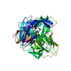
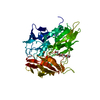
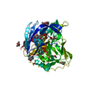
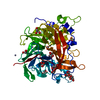
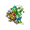
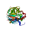
- PDB-1h4p: Crystal structure of exo-1,3-beta glucanse from Saccharomyces cer... -
+ Open data

- Basic information
| Entry | Database: PDB / ID: 1h4p | ||||||||||||
|---|---|---|---|---|---|---|---|---|---|---|---|---|---|
| Title | Crystal structure of exo-1,3-beta glucanse from Saccharomyces cerevisiae | ||||||||||||
 Components Components | GLUCAN 1,3-BETA-GLUCOSIDASE I/II | ||||||||||||
 Keywords Keywords | HYDROLASE / GLUCAN DEGRADATION / HYDROLYASE / GLYCOSIDASE | ||||||||||||
| Function / homology |  Function and homology information Function and homology informationglucan metabolic process / glucan 1,3-beta-glucosidase / glucan exo-1,3-beta-glucosidase activity / fungal-type cell wall organization / glucan catabolic process / fungal-type cell wall / fungal-type vacuole / extracellular region Similarity search - Function | ||||||||||||
| Biological species |  | ||||||||||||
| Method |  X-RAY DIFFRACTION / SYNCHROTRON / MOLECULAR REPLACEMENT / Resolution: 1.75 Å X-RAY DIFFRACTION / SYNCHROTRON / MOLECULAR REPLACEMENT / Resolution: 1.75 Å | ||||||||||||
 Authors Authors | Ferguson, A.D. | ||||||||||||
 Citation Citation | Journal: Nat.Struct.Mol.Biol. / Year: 2004 Title: The Er Protein Folding Sensor Udp-Glucose Glycoprotein:Glucosyltransferase Modifies Substrates Distant to Local Changes in Glycoprotein Conformation. Authors: Taylor, S.C. / Ferguson, A.D. / Bergeron, J.J.M. / Thomas, D.Y. | ||||||||||||
| History |
| ||||||||||||
| Remark 700 | SHEET DETERMINATION METHOD: DSSP THE SHEETS PRESENTED AS "AA" AND "BA" IN EACH CHAIN ON SHEET ... SHEET DETERMINATION METHOD: DSSP THE SHEETS PRESENTED AS "AA" AND "BA" IN EACH CHAIN ON SHEET RECORDS BELOW IS ACTUALLY AN 8-STRANDED BARREL THIS IS REPRESENTED BY A 9-STRANDED SHEET IN WHICH THE FIRST AND LAST STRANDS ARE IDENTICAL. |
- Structure visualization
| Structure viewer | Molecule: MolmilJmol/JSmol |
|---|
- Downloads & links
-Download
| PDBx/mmCIF format | 1h4p.cif.gz | 193.9 KB | Display | PDBx/mmCIF format |
|---|---|---|---|---|
| PDB format | pdb1h4p.ent.gz | 154.8 KB | Display | PDB format |
| PDBx/mmJSON format | 1h4p.json.gz | Tree view | PDBx/mmJSON format | |
| Others |  Other downloads Other downloads |
-Validation report
| Arichive directory | https://data.pdbj.org/pub/pdb/validation_reports/h4/1h4pftp://data.pdbj.org/pub/pdb/validation_reports/h4/1h4p | HTTPS FTP |
|---|
-Related structure data
| Related structure data | 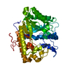 1cz1S S: Starting model for refinement |
|---|---|
| Similar structure data |


-Links
 PDBj
PDBj- Assembly
| Deposited unit | 
| ||||||||
|---|---|---|---|---|---|---|---|---|---|
| 1 | 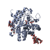
| ||||||||
| 2 | 
| ||||||||
| Unit cell |
| ||||||||
| Noncrystallographic symmetry (NCS) | NCS oper: (Code: given Matrix: (-0.0276, -0.9949, 0.09705), Vector: |
-Components
-Protein , 1 types, 2 molecules AB
| #1: Protein | Mass: 47003.746 Da / Num. of mol.: 2 Source method: isolated from a genetically manipulated source Details: TWO MAN9GLCNAC GLYCAN CHAINS ARE LOCATED AT RESIDUES N165 AND N325 Source: (gene. exp.) Production host: |
|---|
-Sugars , 4 types, 4 molecules
| #2: Polysaccharide | beta-D-mannopyranose-(1-2)-alpha-D-mannopyranose-(1-3)-[alpha-D-mannopyranose-(1-2)-beta-D- ...beta-D-mannopyranose-(1-2)-alpha-D-mannopyranose-(1-3)-[alpha-D-mannopyranose-(1-2)-beta-D-mannopyranose-(1-6)]alpha-D-mannopyranose-(1-6)-[beta-D-mannopyranose-(1-2)-beta-D-mannopyranose-(1-3)]beta-D-mannopyranose-(1-4)-2-acetamido-2-deoxy-beta-D-glucopyranose-(1-4)-2-acetamido-2-deoxy-beta-D-glucopyranose Source method: isolated from a genetically manipulated source |
|---|---|
| #3: Polysaccharide | beta-D-mannopyranose-(1-3)-[beta-D-mannopyranose-(1-6)]beta-D-mannopyranose-(1-4)-2-acetamido-2- ...beta-D-mannopyranose-(1-3)-[beta-D-mannopyranose-(1-6)]beta-D-mannopyranose-(1-4)-2-acetamido-2-deoxy-beta-D-glucopyranose-(1-4)-2-acetamido-2-deoxy-beta-D-glucopyranose Source method: isolated from a genetically manipulated source |
| #4: Polysaccharide | beta-D-mannopyranose-(1-2)-beta-D-mannopyranose-(1-3)-[beta-D-mannopyranose-(1-3)-[beta-D- ...beta-D-mannopyranose-(1-2)-beta-D-mannopyranose-(1-3)-[beta-D-mannopyranose-(1-3)-[beta-D-mannopyranose-(1-6)]beta-D-mannopyranose-(1-6)]alpha-D-mannopyranose-(1-4)-2-acetamido-2-deoxy-beta-D-glucopyranose-(1-4)-2-acetamido-2-deoxy-beta-D-glucopyranose Source method: isolated from a genetically manipulated source |
| #5: Polysaccharide | 2-acetamido-2-deoxy-beta-D-glucopyranose-(1-4)-2-acetamido-2-deoxy-beta-D-glucopyranose Source method: isolated from a genetically manipulated source |
-Non-polymers , 2 types, 465 molecules 

| #6: Chemical | ChemComp-GOL /  Mass: 92.094 Da / Num. of mol.: 9 / Source method: obtained synthetically / Formula: C3H8O3 Mass: 92.094 Da / Num. of mol.: 9 / Source method: obtained synthetically / Formula: C3H8O3#7: Water | ChemComp-HOH / | Mass: 18.015 Da / Num. of mol.: 456 / Source method: isolated from a natural source / Formula: H2O |
|---|
-Details
| Compound details | GLUCANASES| Has protein modification | Y | |
|---|
-Experimental details
-Experiment
| Experiment | Method: X-RAY DIFFRACTION / Number of used crystals: 1 |
|---|
- Sample preparation
| Crystal | Density Matthews: 2.96 Å3/Da / Density % sol: 58 % | |||||||||||||||||||||||||||||||||||
|---|---|---|---|---|---|---|---|---|---|---|---|---|---|---|---|---|---|---|---|---|---|---|---|---|---|---|---|---|---|---|---|---|---|---|---|---|
| Crystal grow | Temperature: 291 K / Method: vapor diffusion, hanging drop / pH: 8 Details: 100 MM SODIUM HEPES PH 7.8, 25 MM MGCL2, 1.3 M TRI-SODIUM CITRATE, 20% GLYCEROL | |||||||||||||||||||||||||||||||||||
| Crystal grow | *PLUS Temperature: 18 ℃ / pH: 8 / Method: vapor diffusion, hanging drop | |||||||||||||||||||||||||||||||||||
| Components of the solutions | *PLUS
|
-Data collection
| Diffraction | Mean temperature: 100 K |
|---|---|
| Diffraction source | Source: SYNCHROTRON / Site: NSLS  / Beamline: X9B / Wavelength: 0.9786 / Beamline: X9B / Wavelength: 0.9786 |
| Detector | Type: MARRESEARCH / Detector: CCD / Date: Jul 15, 2000 |
| Radiation | Protocol: SINGLE WAVELENGTH / Monochromatic (M) / Laue (L): M / Scattering type: x-ray |
| Radiation wavelength | Wavelength: 0.9786 Å / Relative weight: 1 |
| Reflection | Resolution: 1.75→50 Å / Num. obs: 104421 / % possible obs: 95 % / Redundancy: 12 % / Biso Wilson estimate: 21.8 Å2 / Rmerge(I) obs: 0.043 / Net I/σ(I): 37 |
| Reflection shell | Resolution: 1.75→1.86 Å / Redundancy: 4 % / Rmerge(I) obs: 0.433 / Mean I/σ(I) obs: 2.8 / % possible all: 80 |
| Reflection | *PLUS Highest resolution: 1.75 Å / % possible obs: 97.6 % / Num. measured all: 107423 / Rmerge(I) obs: 0.043 |
| Reflection shell | *PLUS % possible obs: 86.9 % / Rmerge(I) obs: 0.443 / Mean I/σ(I) obs: 2.8 |
- Processing
| Software |
| ||||||||||||||||||||||||||||||||||||||||||||||||||||||||||||
|---|---|---|---|---|---|---|---|---|---|---|---|---|---|---|---|---|---|---|---|---|---|---|---|---|---|---|---|---|---|---|---|---|---|---|---|---|---|---|---|---|---|---|---|---|---|---|---|---|---|---|---|---|---|---|---|---|---|---|---|---|---|
| Refinement | Method to determine structure: MOLECULAR REPLACEMENT Starting model: PDB ENTRY 1CZ1 Resolution: 1.75→36.27 Å / Rfactor Rfree error: 0.003 / Data cutoff high absF: 416551.13 / Isotropic thermal model: RESTRAINED / Cross valid method: THROUGHOUT / σ(F): 0
| ||||||||||||||||||||||||||||||||||||||||||||||||||||||||||||
| Solvent computation | Solvent model: FLAT MODEL / Bsol: 51.8131 Å2 / ksol: 0.407808 e/Å3 | ||||||||||||||||||||||||||||||||||||||||||||||||||||||||||||
| Displacement parameters | Biso mean: 28 Å2
| ||||||||||||||||||||||||||||||||||||||||||||||||||||||||||||
| Refine analyze |
| ||||||||||||||||||||||||||||||||||||||||||||||||||||||||||||
| Refinement step | Cycle: LAST / Resolution: 1.75→36.27 Å
| ||||||||||||||||||||||||||||||||||||||||||||||||||||||||||||
| Refine LS restraints |
| ||||||||||||||||||||||||||||||||||||||||||||||||||||||||||||
| LS refinement shell | Resolution: 1.75→1.86 Å / Rfactor Rfree error: 0.011 / Total num. of bins used: 6
| ||||||||||||||||||||||||||||||||||||||||||||||||||||||||||||
| Xplor file |
| ||||||||||||||||||||||||||||||||||||||||||||||||||||||||||||
| Refinement | *PLUS Lowest resolution: 50 Å / % reflection Rfree: 5 % | ||||||||||||||||||||||||||||||||||||||||||||||||||||||||||||
| Solvent computation | *PLUS | ||||||||||||||||||||||||||||||||||||||||||||||||||||||||||||
| Displacement parameters | *PLUS | ||||||||||||||||||||||||||||||||||||||||||||||||||||||||||||
| Refine LS restraints | *PLUS
|
