 Movie
Movie Controller

[English] 日本語
 Yorodumi
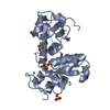
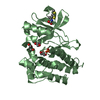
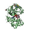
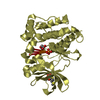
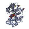
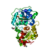
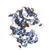
- PDB-1h4k: Sulfurtransferase from Azotobacter vinelandii in complex with hyp... -
+ Open data

- Basic information
| Entry | Database: PDB / ID: 1h4k | ||||||
|---|---|---|---|---|---|---|---|
| Title | Sulfurtransferase from Azotobacter vinelandii in complex with hypophosphite | ||||||
 Components Components | SULFURTRANSFERASE | ||||||
 Keywords Keywords | TRANSFERASE / SULFUR METABOLISM / THIOSULFATE:CYANIDE | ||||||
| Function / homology |  Function and homology information Function and homology informationthiosulfate sulfurtransferase / thiosulfate-cyanide sulfurtransferase activity / cytoplasm Similarity search - Function | ||||||
| Biological species |  AZOTOBACTER VINELANDII (bacteria) AZOTOBACTER VINELANDII (bacteria) | ||||||
| Method |  X-RAY DIFFRACTION / SYNCHROTRON / MOLECULAR REPLACEMENT / Resolution: 2.05 Å X-RAY DIFFRACTION / SYNCHROTRON / MOLECULAR REPLACEMENT / Resolution: 2.05 Å | ||||||
 Authors Authors | Bordo, D. / Forlani, F. / Spallarossa, A. / Colnaghi, R. / Carpen, A. / Pagani, S. / Bolognesi, M. | ||||||
 Citation Citation | Journal: Biol.Chem. / Year: 2001 Title: A Persulfurated Cysteine Promotes Active Site Reactivity in Azotobacter Vinelandii Rhodanse Authors: Bordo, D. / Forlani, F. / Spallarossa, A. / Colnaghi, R. / Carpen, A. / Bolognesi, M. / Pagani, S. #1: Journal: J.Mol.Biol. / Year: 2000Title: The Crystal Structure of a Sulfurtransferase from Azotobacter Vinelandii Highliths the Evolutionary Relationship between the Rhodanese and Phosphatase Enzyme Families Authors: Bordo, D. / Deriu, D. / Colnaghi, R. / Carpen, A. / Pagani, S. / Bolognesi, M. | ||||||
| History |
|
- Structure visualization
| Structure viewer | Molecule: MolmilJmol/JSmol |
|---|
- Downloads & links
-Download
| PDBx/mmCIF format | 1h4k.cif.gz | 69.7 KB | Display | PDBx/mmCIF format |
|---|---|---|---|---|
| PDB format | pdb1h4k.ent.gz | 50.6 KB | Display | PDB format |
| PDBx/mmJSON format | 1h4k.json.gz | Tree view | PDBx/mmJSON format | |
| Others |  Other downloads Other downloads |
-Validation report
| Arichive directory | https://data.pdbj.org/pub/pdb/validation_reports/h4/1h4kftp://data.pdbj.org/pub/pdb/validation_reports/h4/1h4k | HTTPS FTP |
|---|
-Related structure data
| Related structure data |  1h4mC 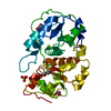 1e0cS S: Starting model for refinement C: citing same article ( |
|---|---|
| Similar structure data |


-Links
 PDBj
PDBj- Assembly
| Deposited unit | 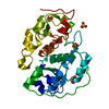
| ||||||||
|---|---|---|---|---|---|---|---|---|---|
| 1 |
| ||||||||
| Unit cell |
|
-Components
| #1: Protein | Mass: 29664.412 Da / Num. of mol.: 1 Source method: isolated from a genetically manipulated source Source: (gene. exp.) AZOTOBACTER VINELANDII (bacteria) / Description: SYNTHETIC GENE / Cellular location: CYTOPLASM / Gene: RHDA / Plasmid: PQE32 / Cellular location (production host): CYTOPLASM / Production host: | ||||||
|---|---|---|---|---|---|---|---|
| #2: Chemical | ChemComp-PO2 /   Mass: 62.973 Da / Num. of mol.: 1 / Source method: obtained synthetically / Formula: O2P Mass: 62.973 Da / Num. of mol.: 1 / Source method: obtained synthetically / Formula: O2P | ||||||
| #3: Chemical |   Mass: 96.063 Da / Num. of mol.: 2 / Source method: obtained synthetically / Formula: SO4 Mass: 96.063 Da / Num. of mol.: 2 / Source method: obtained synthetically / Formula: SO4#4: Chemical |   Mass: 62.068 Da / Num. of mol.: 2 / Source method: obtained synthetically / Formula: C2H6O2 Mass: 62.068 Da / Num. of mol.: 2 / Source method: obtained synthetically / Formula: C2H6O2#5: Water | ChemComp-HOH / |  Mass: 18.015 Da / Num. of mol.: 196 / Source method: isolated from a natural source / Formula: H2O Mass: 18.015 Da / Num. of mol.: 196 / Source method: isolated from a natural source / Formula: H2OCompound details | OTHER_DETAILS: HYPOPHOSPH | |
-Experimental details
-Experiment
| Experiment | Method: X-RAY DIFFRACTION / Number of used crystals: 1 |
|---|
- Sample preparation
| Crystal | Density Matthews: 3 Å3/Da / Density % sol: 50 % |
|---|---|
| Crystal grow | pH: 6.5 / Details: 1.7 M MGSO4, 50 MM MES PH 6.0, 5% (V/V)ETHANEDIOL |
-Data collection
| Diffraction | Mean temperature: 100 K |
|---|---|
| Diffraction source | Source: SYNCHROTRON / Site: EMBL/DESY, HAMBURG  / Beamline: X11 / Wavelength: 0.8349 / Beamline: X11 / Wavelength: 0.8349 |
| Detector | Type: MARRESEARCH |
| Radiation | Protocol: SINGLE WAVELENGTH / Monochromatic (M) / Laue (L): M / Scattering type: x-ray |
| Radiation wavelength | Wavelength: 0.8349 Å / Relative weight: 1 |
| Reflection | Resolution: 2.05→30 Å / Num. obs: 18208 / % possible obs: 92.4 % / Redundancy: 5.7 % / Rmerge(I) obs: 0.094 / Rsym value: 0.094 / Net I/σ(I): 10.5 |
| Reflection shell | Resolution: 2.05→2.09 Å / Redundancy: 2 % / Rmerge(I) obs: 0.3 / Mean I/σ(I) obs: 3.8 / Rsym value: 0.3 / % possible all: 90.2 |
- Processing
| Software |
| ||||||||||||||||||||||||||||||||||||||||||||||||||||||||||||
|---|---|---|---|---|---|---|---|---|---|---|---|---|---|---|---|---|---|---|---|---|---|---|---|---|---|---|---|---|---|---|---|---|---|---|---|---|---|---|---|---|---|---|---|---|---|---|---|---|---|---|---|---|---|---|---|---|---|---|---|---|---|
| Refinement | Method to determine structure: MOLECULAR REPLACEMENT Starting model: PDB ENTRY 1E0C Resolution: 2.05→30 Å / σ(F): 0
| ||||||||||||||||||||||||||||||||||||||||||||||||||||||||||||
| Refinement step | Cycle: LAST / Resolution: 2.05→30 Å
| ||||||||||||||||||||||||||||||||||||||||||||||||||||||||||||
| Refine LS restraints |
| ||||||||||||||||||||||||||||||||||||||||||||||||||||||||||||
| Xplor file | Serial no: 1 / Param file: PARAM19X.PRO |
