 Movie
Movie Controller

[English] 日本語
 Yorodumi
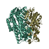
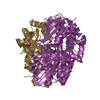
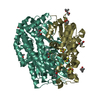
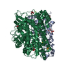
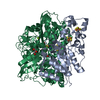
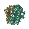
- PDB-1h2r: THREE-DIMENSIONAL STRUCTURE OF NI-FE HYDROGENASE FROM DESULFIVIBR... -
+ Open data

- Basic information
| Entry | Database: PDB / ID: 1h2r | ||||||
|---|---|---|---|---|---|---|---|
| Title | THREE-DIMENSIONAL STRUCTURE OF NI-FE HYDROGENASE FROM DESULFIVIBRIO VULGARIS MIYAZAKI F IN THE REDUCED FORM AT 1.4 A RESOLUTION | ||||||
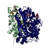
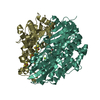
 Components Components | (PROTEIN (PERIPLASMIC [NIFE] HYDROGENASE ...) x 2 | ||||||
 Keywords Keywords | OXIDOREDUCTASE / HIGH RESOLUTION CRYSTAL STRUCTURE / SULFUR-BRIDGING LIGAND / NI-FE HYDROGENASE / REDUCED ENZYME / ATOMIC CAP AT ACTIVE SITE | ||||||
| Function / homology |  Function and homology information Function and homology informationcytochrome-c3 hydrogenase / cytochrome-c3 hydrogenase activity / ferredoxin hydrogenase complex / [Ni-Fe] hydrogenase complex / ferredoxin hydrogenase activity / anaerobic respiration / 3 iron, 4 sulfur cluster binding / nickel cation binding / 4 iron, 4 sulfur cluster binding / periplasmic space ...cytochrome-c3 hydrogenase / cytochrome-c3 hydrogenase activity / ferredoxin hydrogenase complex / [Ni-Fe] hydrogenase complex / ferredoxin hydrogenase activity / anaerobic respiration / 3 iron, 4 sulfur cluster binding / nickel cation binding / 4 iron, 4 sulfur cluster binding / periplasmic space / electron transfer activity / membrane / metal ion binding Similarity search - Function | ||||||
| Biological species |  Desulfovibrio vulgaris str. 'Miyazaki F' (bacteria) Desulfovibrio vulgaris str. 'Miyazaki F' (bacteria) | ||||||
| Method |  X-RAY DIFFRACTION / SYNCHROTRON / D-FOURIER / Resolution: 1.4 Å X-RAY DIFFRACTION / SYNCHROTRON / D-FOURIER / Resolution: 1.4 Å | ||||||
 Authors Authors | Higuchi, Y. / Ogata, H. | ||||||
 Citation Citation | Journal: Structure Fold.Des. / Year: 1999 Title: Removal of the bridging ligand atom at the Ni-Fe active site of [NiFe] hydrogenase upon reduction with H2, as revealed by X-ray structure analysis at 1.4 A resolution. Authors: Higuchi, Y. / Ogata, H. / Miki, K. / Yasuoka, N. / Yagi, T. #1: Journal: Biochem.Biophys.Res.Commun. / Year: 1999Title: Liberation of Hydrogen Sulfide During the Catalytic Action of Desulfoviborio Hydrogenase Under the Atmosphere of Hydrogen Authors: Higuchi, Y. / Yagi, T. #2: Journal: Structure / Year: 1997Title: Unusual Ligand Structure in Ni-Fe Active Center and an Additional Mg Site in Hydrogenase Revealed by High Resolution X-Ray Structure Analysis Authors: Higuchi, Y. / Yagi, T. / Yasuoka, N. | ||||||
| History |
|
- Structure visualization
| Structure viewer | Molecule: MolmilJmol/JSmol |
|---|
- Downloads & links
-Download
| PDBx/mmCIF format | 1h2r.cif.gz | 182.5 KB | Display | PDBx/mmCIF format |
|---|---|---|---|---|
| PDB format | pdb1h2r.ent.gz | 141.5 KB | Display | PDB format |
| PDBx/mmJSON format | 1h2r.json.gz | Tree view | PDBx/mmJSON format | |
| Others |  Other downloads Other downloads |
-Validation report
| Arichive directory | https://data.pdbj.org/pub/pdb/validation_reports/h2/1h2rftp://data.pdbj.org/pub/pdb/validation_reports/h2/1h2r | HTTPS FTP |
|---|
-Related structure data
| Related structure data |  1h2aS S: Starting model for refinement |
|---|---|
| Similar structure data |


-Links
 PDBj
PDBj


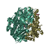
- Assembly
| Deposited unit | 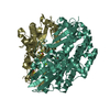
| ||||||||
|---|---|---|---|---|---|---|---|---|---|
| 1 |
| ||||||||
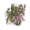
| Unit cell |
|
-Components
-PROTEIN (PERIPLASMIC [NIFE] HYDROGENASE ... , 2 types, 2 molecules SL
| #1: Protein | Mass: 28789.746 Da / Num. of mol.: 1 / Source method: isolated from a natural source Source: (natural) Desulfovibrio vulgaris str. 'Miyazaki F' (bacteria)Cellular location: PERIPLASMIC MEMBRANE / Species: Desulfovibrio vulgaris / Strain: MIYAZAKI F / References: UniProt: P21853, cytochrome-c3 hydrogenase |
|---|---|
| #2: Protein | Mass: 59179.289 Da / Num. of mol.: 1 / Source method: isolated from a natural source Source: (natural) Desulfovibrio vulgaris str. 'Miyazaki F' (bacteria)Cellular location: PERIPLASMIC MEMBRANE / Species: Desulfovibrio vulgaris / Strain: MIYAZAKI F / References: UniProt: P21852, cytochrome-c3 hydrogenase |
-Non-polymers , 5 types, 582 molecules 


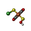

| #3: Chemical |  Mass: 351.640 Da / Num. of mol.: 2 / Source method: obtained synthetically / Formula: Fe4S4 Mass: 351.640 Da / Num. of mol.: 2 / Source method: obtained synthetically / Formula: Fe4S4#4: Chemical | ChemComp-F3S / |  Mass: 295.795 Da / Num. of mol.: 1 / Source method: obtained synthetically / Formula: Fe3S4 Mass: 295.795 Da / Num. of mol.: 1 / Source method: obtained synthetically / Formula: Fe3S4#5: Chemical | ChemComp-MG / |  Mass: 24.305 Da / Num. of mol.: 1 / Source method: obtained synthetically / Formula: Mg Mass: 24.305 Da / Num. of mol.: 1 / Source method: obtained synthetically / Formula: Mg#6: Chemical | ChemComp-NFE / |  Mass: 251.696 Da / Num. of mol.: 1 / Source method: obtained synthetically / Formula: C2HFeNiO3S2 Mass: 251.696 Da / Num. of mol.: 1 / Source method: obtained synthetically / Formula: C2HFeNiO3S2#7: Water | ChemComp-HOH / | Mass: 18.015 Da / Num. of mol.: 577 / Source method: isolated from a natural source / Formula: H2O |
|---|
-Experimental details
-Experiment
| Experiment | Method: X-RAY DIFFRACTION |
|---|
- Sample preparation
| Crystal | Density Matthews: 2.4 Å3/Da / Density % sol: 49 % | |||||||||||||||||||||||||||||||||||||||||||||||||||||||||||||||
|---|---|---|---|---|---|---|---|---|---|---|---|---|---|---|---|---|---|---|---|---|---|---|---|---|---|---|---|---|---|---|---|---|---|---|---|---|---|---|---|---|---|---|---|---|---|---|---|---|---|---|---|---|---|---|---|---|---|---|---|---|---|---|---|---|
| Crystal grow | pH: 7 / Details: 45% MPD, pH 7.0 | |||||||||||||||||||||||||||||||||||||||||||||||||||||||||||||||
| Crystal grow | *PLUS pH: 7.4 / Method: microdialysis / Details: Higuchi, Y., (1987) J.Biol.Chem., 262, 2823. | |||||||||||||||||||||||||||||||||||||||||||||||||||||||||||||||
| Components of the solutions | *PLUS
|
-Data collection
| Diffraction | Mean temperature: 280 K |
|---|---|
| Diffraction source | Source: SYNCHROTRON / Site: SPring-8  / Beamline: BL41XU / Wavelength: 0.708 / Beamline: BL41XU / Wavelength: 0.708 |
| Detector | Detector: IMAGE PLATE |
| Radiation | Protocol: SINGLE WAVELENGTH / Monochromatic (M) / Laue (L): M / Scattering type: x-ray |
| Radiation wavelength | Wavelength: 0.708 Å / Relative weight: 1 |
| Reflection | Resolution: 1.4→20 Å / Num. obs: 155198 / % possible obs: 91.6 % / Observed criterion σ(I): 0 / Redundancy: 9.4 % / Rmerge(I) obs: 0.048 / Rsym value: 0.036 |
| Reflection shell | Resolution: 1.4→1.46 Å / Rmerge(I) obs: 0.38 / Rsym value: 0.3 / % possible all: 85.1 |
| Reflection | *PLUS Num. measured all: 1453748 |
| Reflection shell | *PLUS % possible obs: 85.8 % / Rmerge(I) obs: 0.38 |
- Processing
| Software |
| ||||||||||||||||||||||||||||||||||||||||||||||||||||||||||||
|---|---|---|---|---|---|---|---|---|---|---|---|---|---|---|---|---|---|---|---|---|---|---|---|---|---|---|---|---|---|---|---|---|---|---|---|---|---|---|---|---|---|---|---|---|---|---|---|---|---|---|---|---|---|---|---|---|---|---|---|---|---|
| Refinement | Method to determine structure: D-FOURIER Starting model: 1H2A Resolution: 1.4→6 Å / σ(F): 1
| ||||||||||||||||||||||||||||||||||||||||||||||||||||||||||||
| Displacement parameters | Biso mean: 17.8 Å2 | ||||||||||||||||||||||||||||||||||||||||||||||||||||||||||||
| Refine analyze | Luzzati coordinate error obs: 0.2 Å | ||||||||||||||||||||||||||||||||||||||||||||||||||||||||||||
| Refinement step | Cycle: LAST / Resolution: 1.4→6 Å
| ||||||||||||||||||||||||||||||||||||||||||||||||||||||||||||
| Refine LS restraints |
| ||||||||||||||||||||||||||||||||||||||||||||||||||||||||||||
| LS refinement shell | Resolution: 1.4→1.46 Å / Total num. of bins used: 8
| ||||||||||||||||||||||||||||||||||||||||||||||||||||||||||||
| Software | *PLUS Name: X-PLOR / Classification: refinement | ||||||||||||||||||||||||||||||||||||||||||||||||||||||||||||
| Refinement | *PLUS Highest resolution: 1.4 Å / Lowest resolution: 6 Å / σ(F): 1 / % reflection Rfree: 7 % | ||||||||||||||||||||||||||||||||||||||||||||||||||||||||||||
| Solvent computation | *PLUS | ||||||||||||||||||||||||||||||||||||||||||||||||||||||||||||
| Displacement parameters | *PLUS Biso mean: 17.8 Å2 | ||||||||||||||||||||||||||||||||||||||||||||||||||||||||||||
| LS refinement shell | *PLUS Rfactor Rfree: 0.313 / % reflection Rfree: 7 % / Rfactor Rwork: 0.306 / Rfactor obs: 0.306 |
