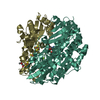
 Movie
Movie Controller


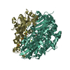
+ Open data

- Basic information
| Entry | Database: PDB / ID: 1h2a | ||||||
|---|---|---|---|---|---|---|---|
| Title | SINGLE CRYSTALS OF HYDROGENASE FROM DESULFOVIBRIO VULGARIS | ||||||
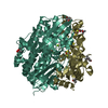
 Components Components | (HYDROGENASE) x 2 | ||||||
 Keywords Keywords | OXIDOREDUCTASE / NI-FE HYDROGENASE / SO LIGAND / HYDROGEN METABOLISM / MG CENTER / MIR / MAD | ||||||
| Function / homology |  Function and homology information Function and homology informationcytochrome-c3 hydrogenase / cytochrome-c3 hydrogenase activity / ferredoxin hydrogenase complex / [Ni-Fe] hydrogenase complex / ferredoxin hydrogenase activity / anaerobic respiration / 3 iron, 4 sulfur cluster binding / nickel cation binding / 4 iron, 4 sulfur cluster binding / periplasmic space ...cytochrome-c3 hydrogenase / cytochrome-c3 hydrogenase activity / ferredoxin hydrogenase complex / [Ni-Fe] hydrogenase complex / ferredoxin hydrogenase activity / anaerobic respiration / 3 iron, 4 sulfur cluster binding / nickel cation binding / 4 iron, 4 sulfur cluster binding / periplasmic space / electron transfer activity / membrane / metal ion binding Similarity search - Function | ||||||
| Biological species |  Desulfovibrio vulgaris str. 'Miyazaki F' (bacteria) Desulfovibrio vulgaris str. 'Miyazaki F' (bacteria) | ||||||
| Method |  X-RAY DIFFRACTION / SYNCHROTRON / MIR, MAD / Resolution: 1.8 Å X-RAY DIFFRACTION / SYNCHROTRON / MIR, MAD / Resolution: 1.8 Å | ||||||
 Authors Authors | Higuchi, Y. / Yasuoka, N. | ||||||
 Citation Citation | Journal: Structure / Year: 1997 Title: Unusual ligand structure in Ni-Fe active center and an additional Mg site in hydrogenase revealed by high resolution X-ray structure analysis. Authors: Higuchi, Y. / Yagi, T. / Yasuoka, N. #1: Journal: Acta Crystallogr.,Sect.D / Year: 1994Title: Location of Active Sites of Nife Hydrogenase Determined by the Combination of Multiple Isomorphous Replacement and Multiwavelength Anomalous-Diffraction Methods Authors: Higuchi, Y. / Okamoto, T. / Fujimoto, K. / Misaki, S. / Morimoto, Y. / Yasouka, N. #2: Journal: J.Biol.Chem. / Year: 1987Title: Single Crystals of Hydrogenase from Desulfovibrio Vulgaris Miyazaki F Authors: Higuchi, Y. / Yasuoka, N. / Kakudo, M. / Katsube, Y. / Yagi, T. / Inokuchi, H. | ||||||
| History |
|
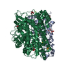
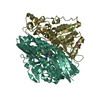
- Structure visualization
| Structure viewer | Molecule: MolmilJmol/JSmol |
|---|
- Downloads & links
-Download
| PDBx/mmCIF format | 1h2a.cif.gz | 180.4 KB | Display | PDBx/mmCIF format |
|---|---|---|---|---|
| PDB format | pdb1h2a.ent.gz | 138.7 KB | Display | PDB format |
| PDBx/mmJSON format | 1h2a.json.gz | Tree view | PDBx/mmJSON format | |
| Others |  Other downloads Other downloads |
-Validation report
| Arichive directory | https://data.pdbj.org/pub/pdb/validation_reports/h2/1h2aftp://data.pdbj.org/pub/pdb/validation_reports/h2/1h2a | HTTPS FTP |
|---|
-Related structure data
| Similar structure data |
|---|

-Links
 PDBj
PDBj


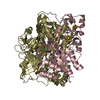
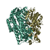
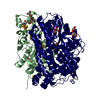
- Assembly
| Deposited unit | 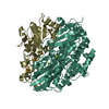
| ||||||||
|---|---|---|---|---|---|---|---|---|---|
| 1 |
| ||||||||
| Unit cell |
|
-Components
-Protein , 2 types, 2 molecules SL
| #1: Protein | Mass: 34148.043 Da / Num. of mol.: 1 / Source method: isolated from a natural source / Details: IAM 12604 Source: (natural) Desulfovibrio vulgaris str. 'Miyazaki F' (bacteria)Species: Desulfovibrio vulgaris / Strain: MIYAZAKI F / References: UniProt: P21853, 1.18.99.1 |
|---|---|
| #2: Protein | Mass: 62711.422 Da / Num. of mol.: 1 / Source method: isolated from a natural source / Details: IAM 12604 Source: (natural) Desulfovibrio vulgaris str. 'Miyazaki F' (bacteria)Species: Desulfovibrio vulgaris / Strain: MIYAZAKI F / References: UniProt: P21852, 1.18.99.1 |
-Non-polymers , 5 types, 643 molecules 


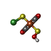

| #3: Chemical |  Mass: 351.640 Da / Num. of mol.: 2 / Source method: obtained synthetically / Formula: Fe4S4 Mass: 351.640 Da / Num. of mol.: 2 / Source method: obtained synthetically / Formula: Fe4S4#4: Chemical | ChemComp-F3S / |  Mass: 295.795 Da / Num. of mol.: 1 / Source method: obtained synthetically / Formula: Fe3S4 Mass: 295.795 Da / Num. of mol.: 1 / Source method: obtained synthetically / Formula: Fe3S4#5: Chemical | ChemComp-MG / |  Mass: 24.305 Da / Num. of mol.: 1 / Source method: obtained synthetically / Formula: Mg Mass: 24.305 Da / Num. of mol.: 1 / Source method: obtained synthetically / Formula: Mg#6: Chemical | ChemComp-NFE / |  Mass: 251.696 Da / Num. of mol.: 1 / Source method: obtained synthetically / Formula: C2HFeNiO3S2 Mass: 251.696 Da / Num. of mol.: 1 / Source method: obtained synthetically / Formula: C2HFeNiO3S2#7: Water | ChemComp-HOH / | Mass: 18.015 Da / Num. of mol.: 638 / Source method: isolated from a natural source / Formula: H2O |
|---|
-Experimental details
-Experiment
| Experiment | Method: X-RAY DIFFRACTION |
|---|
- Sample preparation
| Crystal | Density Matthews: 2.4 Å3/Da / Density % sol: 49 % | |||||||||||||||||||||||||||||||||||||||||||||||||||||||||||||||
|---|---|---|---|---|---|---|---|---|---|---|---|---|---|---|---|---|---|---|---|---|---|---|---|---|---|---|---|---|---|---|---|---|---|---|---|---|---|---|---|---|---|---|---|---|---|---|---|---|---|---|---|---|---|---|---|---|---|---|---|---|---|---|---|---|
| Crystal grow | pH: 7 / Details: pH 7.0 | |||||||||||||||||||||||||||||||||||||||||||||||||||||||||||||||
| Crystal grow | *PLUS pH: 7.4 / Method: microdialysis / Details: Higuchi, Y., (1987) J.Biol.Chem., 262, 2823. | |||||||||||||||||||||||||||||||||||||||||||||||||||||||||||||||
| Components of the solutions | *PLUS
|
-Data collection
| Diffraction | Mean temperature: 280 K |
|---|---|
| Diffraction source | Source: SYNCHROTRON / Site: Photon Factory  / Beamline: BL-6A / Wavelength: 1 / Beamline: BL-6A / Wavelength: 1 |
| Detector | Detector: IMAGE PLATE |
| Radiation | Monochromator: SI(111) / Monochromatic (M) / Laue (L): M / Scattering type: x-ray |
| Radiation wavelength | Wavelength: 1 Å / Relative weight: 1 |
| Reflection | Resolution: 1.8→20 Å / Num. obs: 251414 / % possible obs: 81.3 % / Observed criterion σ(I): 0 / Redundancy: 4 % / Rmerge(I) obs: 0.105 / Rsym value: 0.041 |
| Reflection shell | Resolution: 1.8→1.88 Å / % possible all: 46 |
| Reflection | *PLUS Num. obs: 63133 / Num. measured all: 251414 |
| Reflection shell | *PLUS % possible obs: 46 % |
- Processing
| Software |
| ||||||||||||||||||||||||||||||||||||||||||||||||||||||||||||
|---|---|---|---|---|---|---|---|---|---|---|---|---|---|---|---|---|---|---|---|---|---|---|---|---|---|---|---|---|---|---|---|---|---|---|---|---|---|---|---|---|---|---|---|---|---|---|---|---|---|---|---|---|---|---|---|---|---|---|---|---|---|
| Refinement | Method to determine structure: MIR, MAD / Resolution: 1.8→20 Å / σ(F): 1
| ||||||||||||||||||||||||||||||||||||||||||||||||||||||||||||
| Displacement parameters | Biso mean: 24.3 Å2 | ||||||||||||||||||||||||||||||||||||||||||||||||||||||||||||
| Refine analyze | Luzzati coordinate error obs: 0.3 Å | ||||||||||||||||||||||||||||||||||||||||||||||||||||||||||||
| Refinement step | Cycle: LAST / Resolution: 1.8→20 Å
| ||||||||||||||||||||||||||||||||||||||||||||||||||||||||||||
| Refine LS restraints |
| ||||||||||||||||||||||||||||||||||||||||||||||||||||||||||||
| Software | *PLUS Name: X-PLOR / Classification: refinement | ||||||||||||||||||||||||||||||||||||||||||||||||||||||||||||
| Refinement | *PLUS | ||||||||||||||||||||||||||||||||||||||||||||||||||||||||||||
| Solvent computation | *PLUS | ||||||||||||||||||||||||||||||||||||||||||||||||||||||||||||
| Displacement parameters | *PLUS | ||||||||||||||||||||||||||||||||||||||||||||||||||||||||||||
| Refine LS restraints | *PLUS
|
