 Movie
Movie Controller


+ Open data

- Basic information
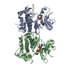
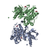
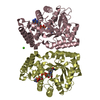
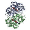
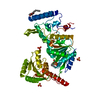
| Entry | Database: PDB / ID: 1gve | ||||||
|---|---|---|---|---|---|---|---|
| Title | Aflatoxin aldehyde reductase (AKR7A1) from Rat Liver | ||||||
 Components Components | AFLATOXIN B1 ALDEHYDE REDUCTASE MEMBER 3 | ||||||
 Keywords Keywords | OXIDOREDUCTASE / ALDO-KETO REDUCTASE / AFLATOXIN B1 / SUCCINIC SEMIALDEHYDE OXIDOREDUCTASE / AKR7 FAMILY | ||||||
| Function / homology |  Function and homology information Function and homology informationAflatoxin activation and detoxification / aflatoxin catabolic process / alcohol dehydrogenase (NADP+) / oxidoreductase activity, acting on CH-OH group of donors / aflatoxin metabolic process / alcohol dehydrogenase (NAD+) activity / alcohol dehydrogenase (NADP+) activity / NADP+ binding / Golgi apparatus / identical protein binding ...Aflatoxin activation and detoxification / aflatoxin catabolic process / alcohol dehydrogenase (NADP+) / oxidoreductase activity, acting on CH-OH group of donors / aflatoxin metabolic process / alcohol dehydrogenase (NAD+) activity / alcohol dehydrogenase (NADP+) activity / NADP+ binding / Golgi apparatus / identical protein binding / cytoplasm / cytosol Similarity search - Function | ||||||
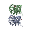
| Biological species |  | ||||||
| Method |  X-RAY DIFFRACTION / SYNCHROTRON / MOLECULAR REPLACEMENT / Resolution: 1.38 Å X-RAY DIFFRACTION / SYNCHROTRON / MOLECULAR REPLACEMENT / Resolution: 1.38 Å | ||||||
 Authors Authors | Kozma, E. / Brown, E. / Ellis, E.M. / Lapthorn, A.J. | ||||||
 Citation Citation | Journal: J.Biol.Chem. / Year: 2002 Title: The Crystal Structure of Rat Liver Akr7A1: A Dimeric Member of the Aldo-Keto Reductase Superfamily Authors: Kozma, E. / Brown, E. / Ellis, E.M. / Lapthorn, A.J. #1: Journal: Proc.Natl.Acad.Sci.USA / Year: 1993 Title: An Ethoxyquin-Inducible Aldehyde Reductase from Rat Liver that Metabolizes Aflatoxin B1 Defines a Subfamily of Aldo-Keto Reductases Authors: Ellis, E.M. / Judah, D.J. / Neal, G.E. / Hayes, J.D. | ||||||
| History |
| ||||||
| Remark 700 | SHEET DETERMINATION METHOD: DSSP THE SHEETS PRESENTED AS "AA" IN EACH CHAIN ON SHEET RECORDS BELOW ... SHEET DETERMINATION METHOD: DSSP THE SHEETS PRESENTED AS "AA" IN EACH CHAIN ON SHEET RECORDS BELOW IS ACTUALLY AN 9-STRANDED BARREL THIS IS REPRESENTED BY A 10-STRANDED SHEET IN WHICH THE FIRST AND LAST STRANDS ARE IDENTICAL. THE SHEETS PRESENTED AS "BA" IN EACH CHAIN ON SHEET RECORDS BELOW IS ACTUALLY AN 9-STRANDED BARREL THIS IS REPRESENTED BY A 10-STRANDED SHEET IN WHICH THE FIRST AND LAST STRANDS ARE IDENTICAL. |
- Structure visualization
| Structure viewer | Molecule: MolmilJmol/JSmol |
|---|
- Downloads & links
-Download
| PDBx/mmCIF format | 1gve.cif.gz | 275.1 KB | Display | PDBx/mmCIF format |
|---|---|---|---|---|
| PDB format | pdb1gve.ent.gz | 222.1 KB | Display | PDB format |
| PDBx/mmJSON format | 1gve.json.gz | Tree view | PDBx/mmJSON format | |
| Others |  Other downloads Other downloads |
-Validation report
| Arichive directory | https://data.pdbj.org/pub/pdb/validation_reports/gv/1gveftp://data.pdbj.org/pub/pdb/validation_reports/gv/1gve | HTTPS FTP |
|---|
-Related structure data
| Related structure data | 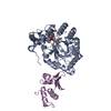 1exbS S: Starting model for refinement |
|---|---|
| Similar structure data |

-Links
 PDBj
PDBj
- Assembly
| Deposited unit | 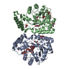
| ||||||||
|---|---|---|---|---|---|---|---|---|---|
| 1 |
| ||||||||
| Unit cell |
| ||||||||
| Noncrystallographic symmetry (NCS) | NCS oper: (Code: given Matrix: (0.97152, -0.04903, -0.23181), Vector: |
-Components
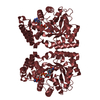
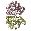
| #1: Protein | Mass: 36789.996 Da / Num. of mol.: 2 Source method: isolated from a genetically manipulated source Source: (gene. exp.)  #2: Chemical | ChemComp-NAP / |   Mass: 743.405 Da / Num. of mol.: 1 / Source method: obtained synthetically / Formula: C21H28N7O17P3 Mass: 743.405 Da / Num. of mol.: 1 / Source method: obtained synthetically / Formula: C21H28N7O17P3#3: Chemical | ChemComp-GOL /   Mass: 92.094 Da / Num. of mol.: 12 / Source method: obtained synthetically / Formula: C3H8O3 Mass: 92.094 Da / Num. of mol.: 12 / Source method: obtained synthetically / Formula: C3H8O3#4: Chemical | ChemComp-CIT / |   Mass: 192.124 Da / Num. of mol.: 1 / Source method: obtained synthetically / Formula: C6H8O7 Mass: 192.124 Da / Num. of mol.: 1 / Source method: obtained synthetically / Formula: C6H8O7#5: Water | ChemComp-HOH / |  Mass: 18.015 Da / Num. of mol.: 504 / Source method: isolated from a natural source / Formula: H2O Mass: 18.015 Da / Num. of mol.: 504 / Source method: isolated from a natural source / Formula: H2O |
|---|
-Experimental details
-Experiment
| Experiment | Method: X-RAY DIFFRACTION / Number of used crystals: 1 |
|---|
- Sample preparation
| Crystal | Density Matthews: 2.7 Å3/Da / Density % sol: 55 % Description: DATA EFFECTIVELY 100% COMPLETE TO 1.7A RESOLUTION. DATA WERE ANISOTROPIC RESULTING IN THE LOW COMPLETENESS AT HIGH RESOLUTION | ||||||||||||||||||||||||
|---|---|---|---|---|---|---|---|---|---|---|---|---|---|---|---|---|---|---|---|---|---|---|---|---|---|
| Crystal grow | Method: vapor diffusion, sitting drop / pH: 5.6 Details: SITTING DROP METHOD WITH 1MICROLITRE PROTEIN (6 MG/ML) AND 1MICROLITRE WELL 20% PEG8K, 0.2M LITHIUM SULPHATE, 0.1M SODIUM CITRATE PH 5. | ||||||||||||||||||||||||
| Crystal grow | *PLUS Temperature: 20 ℃ / Method: vapor diffusion, sitting drop | ||||||||||||||||||||||||
| Components of the solutions | *PLUS
|
-Data collection
| Diffraction | Mean temperature: 100 K |
|---|---|
| Diffraction source | Source: SYNCHROTRON / Site: EMBL/DESY, HAMBURG  / Beamline: BW7B / Wavelength: 0.83 / Beamline: BW7B / Wavelength: 0.83 |
| Detector | Type: MAR scanner 345 mm plate / Detector: IMAGE PLATE / Date: Aug 15, 2000 / Details: BENT MIRROR |
| Radiation | Monochromator: TRIANGULAR / Protocol: SINGLE WAVELENGTH / Monochromatic (M) / Laue (L): M / Scattering type: x-ray |
| Radiation wavelength | Wavelength: 0.83 Å / Relative weight: 1 |
| Reflection | Resolution: 1.38→30 Å / Num. obs: 134602 / % possible obs: 85.4 % / Redundancy: 2.8 % / Rmerge(I) obs: 0.046 / Net I/σ(I): 24 |
| Reflection shell | Resolution: 1.38→1.43 Å / Redundancy: 2.8 % / Rmerge(I) obs: 0.467 / Mean I/σ(I) obs: 1.2 / % possible all: 43 |
| Reflection | *PLUS Num. obs: 158665 |
| Reflection shell | *PLUS % possible obs: 42.8 % |
- Processing
| Software |
| ||||||||||||||||||||||||||||||||||||||||||||||||||||||||||||||||||||||||||||||||||||||||||||||||||||||||||||||||||||||||||||||||||||||||||||||||||||||||||||||||||||||||||||||||||||||
|---|---|---|---|---|---|---|---|---|---|---|---|---|---|---|---|---|---|---|---|---|---|---|---|---|---|---|---|---|---|---|---|---|---|---|---|---|---|---|---|---|---|---|---|---|---|---|---|---|---|---|---|---|---|---|---|---|---|---|---|---|---|---|---|---|---|---|---|---|---|---|---|---|---|---|---|---|---|---|---|---|---|---|---|---|---|---|---|---|---|---|---|---|---|---|---|---|---|---|---|---|---|---|---|---|---|---|---|---|---|---|---|---|---|---|---|---|---|---|---|---|---|---|---|---|---|---|---|---|---|---|---|---|---|---|---|---|---|---|---|---|---|---|---|---|---|---|---|---|---|---|---|---|---|---|---|---|---|---|---|---|---|---|---|---|---|---|---|---|---|---|---|---|---|---|---|---|---|---|---|---|---|---|---|
| Refinement | Method to determine structure: MOLECULAR REPLACEMENT Starting model: PDB ENTRY 1EXB Resolution: 1.38→28.75 Å / Cor.coef. Fo:Fc: 0.973 / Cor.coef. Fo:Fc free: 0.968 / SU B: 2.993 / SU ML: 0.061 / Cross valid method: THROUGHOUT / ESU R: 0.059 / ESU R Free: 0.054 / Stereochemistry target values: MAXIMUM LIKELIHOOD / Details: HYDROGENS HAVE BEEN ADDED IN THE RIDING POSITIONS
| ||||||||||||||||||||||||||||||||||||||||||||||||||||||||||||||||||||||||||||||||||||||||||||||||||||||||||||||||||||||||||||||||||||||||||||||||||||||||||||||||||||||||||||||||||||||
| Solvent computation | Ion probe radii: 0.8 Å / Shrinkage radii: 0.8 Å / VDW probe radii: 1.4 Å / Solvent model: BABINET MODEL WITH MASK | ||||||||||||||||||||||||||||||||||||||||||||||||||||||||||||||||||||||||||||||||||||||||||||||||||||||||||||||||||||||||||||||||||||||||||||||||||||||||||||||||||||||||||||||||||||||
| Refinement step | Cycle: LAST / Resolution: 1.38→28.75 Å
| ||||||||||||||||||||||||||||||||||||||||||||||||||||||||||||||||||||||||||||||||||||||||||||||||||||||||||||||||||||||||||||||||||||||||||||||||||||||||||||||||||||||||||||||||||||||
| Refine LS restraints |
|