 Movie
Movie Controller

[English] 日本語
 Yorodumi
- PDB-1gu6: Structure of the Periplasmic Cytochrome c Nitrite Reductase from ... -
+ Open data

- Basic information
| Entry | Database: PDB / ID: 1gu6 | ||||||
|---|---|---|---|---|---|---|---|
| Title | Structure of the Periplasmic Cytochrome c Nitrite Reductase from Escherichia coli | ||||||
 Components Components | CYTOCHROME C552 | ||||||
 Keywords Keywords | OXIDOREDUCTASE / PERIPLASMIC NITRITE REDUCTASE / C-TYPE CYTOCHROME / ANAEROBIC NITRITE RESPIRATION | ||||||
| Function / homology |  Function and homology information Function and homology informationnitrite reductase (cytochrome; ammonia-forming) / nitrite reductase (cytochrome, ammonia-forming) activity / nitric oxide reductase activity / anaerobic electron transport chain / nitrate assimilation / outer membrane-bounded periplasmic space / iron ion binding / heme binding / calcium ion binding Similarity search - Function | ||||||
| Biological species |  | ||||||
| Method |  X-RAY DIFFRACTION / SYNCHROTRON / MOLECULAR REPLACEMENT / Resolution: 2.5 Å X-RAY DIFFRACTION / SYNCHROTRON / MOLECULAR REPLACEMENT / Resolution: 2.5 Å | ||||||
 Authors Authors | Bamford, V.A. / Angove, H.C. / Seward, H.E. / Thomson, A.J. / Cole, J.A. / Butt, J.N. / Hemmings, A.M. / Richardson, D.J. | ||||||
 Citation Citation | Journal: Biochemistry / Year: 2002 Title: Structure and Spectroscopy of the Periplasmic Cytochrome C Nitrite Reductase from Escherichia Coli Authors: Bamford, V.A. / Angove, H.C. / Seward, H.E. / Thomson, A.J. / Cole, J.A. / Butt, J.N. / Hemmings, A.M. / Richardson, D.J. | ||||||
| History |
|
- Structure visualization
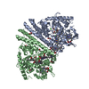
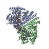
| Structure viewer | Molecule: MolmilJmol/JSmol |
|---|
- Downloads & links
-Download
| PDBx/mmCIF format | 1gu6.cif.gz | 389.7 KB | Display | PDBx/mmCIF format |
|---|---|---|---|---|
| PDB format | pdb1gu6.ent.gz | 322.5 KB | Display | PDB format |
| PDBx/mmJSON format | 1gu6.json.gz | Tree view | PDBx/mmJSON format | |
| Others |  Other downloads Other downloads |
-Validation report
| Arichive directory | https://data.pdbj.org/pub/pdb/validation_reports/gu/1gu6ftp://data.pdbj.org/pub/pdb/validation_reports/gu/1gu6 | HTTPS FTP |
|---|
-Related structure data
| Related structure data |  1qdbS S: Starting model for refinement |
|---|---|
| Similar structure data |
-Links
 PDBj
PDBj


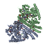
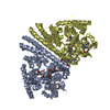
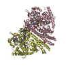
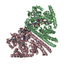
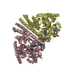
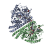
- Assembly
| Deposited unit | 
| ||||||||||||||||
|---|---|---|---|---|---|---|---|---|---|---|---|---|---|---|---|---|---|
| 1 | 
| ||||||||||||||||
| 2 | 
| ||||||||||||||||
| Unit cell |
| ||||||||||||||||
| Noncrystallographic symmetry (NCS) | NCS oper:
|
-Components
| #1: Protein | Mass: 50668.117 Da / Num. of mol.: 4 / Source method: isolated from a natural source / Source: (natural) #2: Chemical | ChemComp-CA /   Mass: 40.078 Da / Num. of mol.: 8 / Source method: obtained synthetically / Formula: Ca Mass: 40.078 Da / Num. of mol.: 8 / Source method: obtained synthetically / Formula: Ca#3: Chemical | ChemComp-GOL /   Mass: 92.094 Da / Num. of mol.: 4 / Source method: obtained synthetically / Formula: C3H8O3 Mass: 92.094 Da / Num. of mol.: 4 / Source method: obtained synthetically / Formula: C3H8O3#4: Chemical | ChemComp-HEC /   Mass: 618.503 Da / Num. of mol.: 20 / Source method: obtained synthetically / Formula: C34H34FeN4O4 Mass: 618.503 Da / Num. of mol.: 20 / Source method: obtained synthetically / Formula: C34H34FeN4O4#5: Water | ChemComp-HOH / |  Mass: 18.015 Da / Num. of mol.: 678 / Source method: isolated from a natural source / Formula: H2O Mass: 18.015 Da / Num. of mol.: 678 / Source method: isolated from a natural source / Formula: H2OCompound details | RESIDUES NUMBERED FROM START OF TRANSLATED | Has protein modification | Y | |
|---|
-Experimental details
-Experiment
| Experiment | Method: X-RAY DIFFRACTION / Number of used crystals: 1 |
|---|
- Sample preparation
| Crystal | Density Matthews: 2.68 Å3/Da / Density % sol: 54.15 % | |||||||||||||||||||||||||||||||||||||||||||||||||
|---|---|---|---|---|---|---|---|---|---|---|---|---|---|---|---|---|---|---|---|---|---|---|---|---|---|---|---|---|---|---|---|---|---|---|---|---|---|---|---|---|---|---|---|---|---|---|---|---|---|---|
| Crystal grow | pH: 7.5 / Details: pH 7.50 | |||||||||||||||||||||||||||||||||||||||||||||||||
| Crystal grow | *PLUS pH: 7 / Method: vapor diffusion | |||||||||||||||||||||||||||||||||||||||||||||||||
| Components of the solutions | *PLUS
|
-Data collection
| Diffraction | Mean temperature: 100 K |
|---|---|
| Diffraction source | Source: SYNCHROTRON / Site: ESRF  / Beamline: ID14-1 / Wavelength: 0.934 / Beamline: ID14-1 / Wavelength: 0.934 |
| Detector | Type: MARRESEARCH / Detector: CCD / Date: Feb 15, 2001 |
| Radiation | Protocol: SINGLE WAVELENGTH / Monochromatic (M) / Laue (L): M / Scattering type: x-ray |
| Radiation wavelength | Wavelength: 0.934 Å / Relative weight: 1 |
| Reflection | Resolution: 2.5→20 Å / Num. obs: 71769 / % possible obs: 93 % / Observed criterion σ(I): 0 / Redundancy: 3.5 % / Rmerge(I) obs: 0.089 / Net I/σ(I): 5.4 |
| Reflection shell | Redundancy: 2.7 % / Rmerge(I) obs: 0.244 / Mean I/σ(I) obs: 2.7 / % possible all: 94.6 |
| Reflection | *PLUS Lowest resolution: 20 Å / % possible obs: 93 % / Num. measured all: 1062813 |
| Reflection shell | *PLUS % possible obs: 94.6 % |
- Processing
| Software |
| ||||||||||||||||||||||||||||||||||||||||||||||||||||||||||||
|---|---|---|---|---|---|---|---|---|---|---|---|---|---|---|---|---|---|---|---|---|---|---|---|---|---|---|---|---|---|---|---|---|---|---|---|---|---|---|---|---|---|---|---|---|---|---|---|---|---|---|---|---|---|---|---|---|---|---|---|---|---|
| Refinement | Method to determine structure: MOLECULAR REPLACEMENT Starting model: 1QDB Resolution: 2.5→20 Å / Cross valid method: THROUGHOUT / σ(F): 0
| ||||||||||||||||||||||||||||||||||||||||||||||||||||||||||||
| Refinement step | Cycle: LAST / Resolution: 2.5→20 Å
| ||||||||||||||||||||||||||||||||||||||||||||||||||||||||||||
| Refine LS restraints |
| ||||||||||||||||||||||||||||||||||||||||||||||||||||||||||||
| Refinement | *PLUS Lowest resolution: 20 Å | ||||||||||||||||||||||||||||||||||||||||||||||||||||||||||||
| Solvent computation | *PLUS | ||||||||||||||||||||||||||||||||||||||||||||||||||||||||||||
| Displacement parameters | *PLUS | ||||||||||||||||||||||||||||||||||||||||||||||||||||||||||||
| LS refinement shell | *PLUS % reflection Rfree: 5 % / Rfactor Rwork: 0.28 / Rfactor obs: 0.28 |
