 Movie
Movie Controller

[English] 日本語
 Yorodumi
- PDB-1gti: MODIFIED GLUTATHIONE S-TRANSFERASE (PI) COMPLEXED WITH S (P-NITRO... -
+ Open data

- Basic information
| Entry | Database: PDB / ID: 1gti | ||||||
|---|---|---|---|---|---|---|---|
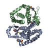
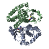
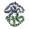
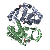
| Title | MODIFIED GLUTATHIONE S-TRANSFERASE (PI) COMPLEXED WITH S (P-NITROBENZYL)GLUTATHIONE | ||||||
 Components Components | GLUTATHIONE S-TRANSFERASE | ||||||
 Keywords Keywords | TRANSFERASE / GLUTATHIONE | ||||||
| Function / homology |  Function and homology information Function and homology informationnegative regulation of neutrophil aggregation / Paracetamol ADME / Glutathione conjugation / Detoxification of Reactive Oxygen Species / S-nitrosoglutathione binding / TRAF2-GSTP1 complex / negative regulation of smooth muscle cell chemotaxis / negative regulation of leukocyte proliferation / dinitrosyl-iron complex binding / common myeloid progenitor cell proliferation ...negative regulation of neutrophil aggregation / Paracetamol ADME / Glutathione conjugation / Detoxification of Reactive Oxygen Species / S-nitrosoglutathione binding / TRAF2-GSTP1 complex / negative regulation of smooth muscle cell chemotaxis / negative regulation of leukocyte proliferation / dinitrosyl-iron complex binding / common myeloid progenitor cell proliferation / hepoxilin biosynthetic process / cellular response to cell-matrix adhesion / glutathione derivative biosynthetic process / response to L-ascorbic acid / linoleic acid metabolic process / negative regulation of monocyte chemotactic protein-1 production / JUN kinase binding / glutathione peroxidase activity / oligodendrocyte development / negative regulation of stress-activated MAPK cascade / negative regulation of JNK cascade / cellular response to glucocorticoid stimulus / prostaglandin metabolic process / negative regulation of interleukin-1 beta production / regulation of stress-activated MAPK cascade / negative regulation of acute inflammatory response / glutathione transferase / negative regulation of ferroptosis / negative regulation of vascular associated smooth muscle cell proliferation / response to amino acid / glutathione transferase activity / animal organ regeneration / negative regulation of tumor necrosis factor production / protein serine/threonine kinase inhibitor activity / regulation of ERK1 and ERK2 cascade / toxic substance binding / negative regulation of fibroblast proliferation / positive regulation of superoxide anion generation / Neutrophil degranulation / xenobiotic metabolic process / cellular response to epidermal growth factor stimulus / fatty acid binding / negative regulation of extrinsic apoptotic signaling pathway / response to reactive oxygen species / negative regulation of canonical NF-kappaB signal transduction / glutathione metabolic process / response to nutrient levels / negative regulation of ERK1 and ERK2 cascade / response to toxic substance / cellular response to insulin stimulus / response to estradiol / cellular response to lipopolysaccharide / response to ethanol / inflammatory response / protein kinase binding / negative regulation of apoptotic process / negative regulation of transcription by RNA polymerase II / protein-containing complex / mitochondrion / nucleus / cytosol Similarity search - Function | ||||||
| Biological species |  | ||||||
| Method |  X-RAY DIFFRACTION / SYNCHROTRON / MOLECULAR REPLACEMENT / Resolution: 3 Å X-RAY DIFFRACTION / SYNCHROTRON / MOLECULAR REPLACEMENT / Resolution: 3 Å | ||||||
 Authors Authors | Vega, M.C. / Coll, M. | ||||||
 Citation Citation | Journal: J.Biol.Chem. / Year: 1998 Title: The three-dimensional structure of Cys-47-modified mouse liver glutathione S-transferase P1-1. Carboxymethylation dramatically decreases the affinity for glutathione and is associated with a ...Title: The three-dimensional structure of Cys-47-modified mouse liver glutathione S-transferase P1-1. Carboxymethylation dramatically decreases the affinity for glutathione and is associated with a loss of electron density in the alphaB-310B region. Authors: Vega, M.C. / Walsh, S.B. / Mantle, T.J. / Coll, M. #1: Journal: J.Mol.Biol. / Year: 1994Title: Molecular Structure at 1.8 A of Mouse Liver Class Pi Glutathione S-Transferase Complexed with S-(P-Nitrobenzyl)Glutathione and Other Inhibitors Authors: Garcia-Saez, I. / Parraga, A. / Phillips, M.F. / Mantle, T.J. / Coll, M. | ||||||
| History |
|
- Structure visualization
| Structure viewer | Molecule: MolmilJmol/JSmol |
|---|
- Downloads & links
-Download
| PDBx/mmCIF format | 1gti.cif.gz | 247.5 KB | Display | PDBx/mmCIF format |
|---|---|---|---|---|
| PDB format | pdb1gti.ent.gz | 203.7 KB | Display | PDB format |
| PDBx/mmJSON format | 1gti.json.gz | Tree view | PDBx/mmJSON format | |
| Others |  Other downloads Other downloads |
-Validation report
| Arichive directory | https://data.pdbj.org/pub/pdb/validation_reports/gt/1gtiftp://data.pdbj.org/pub/pdb/validation_reports/gt/1gti | HTTPS FTP |
|---|
-Related structure data
| Related structure data | 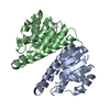 1bayC 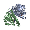 1glqS S: Starting model for refinement C: citing same article ( |
|---|---|
| Similar structure data |


-Links
 PDBj
PDBj


- Assembly
| Deposited unit | 
| |||||||||||||||||||||||||||||||||
|---|---|---|---|---|---|---|---|---|---|---|---|---|---|---|---|---|---|---|---|---|---|---|---|---|---|---|---|---|---|---|---|---|---|---|
| 1 | 
| |||||||||||||||||||||||||||||||||
| 2 | 
| |||||||||||||||||||||||||||||||||
| 3 | 
| |||||||||||||||||||||||||||||||||
| Unit cell |
| |||||||||||||||||||||||||||||||||
| Noncrystallographic symmetry (NCS) | NCS domain:
NCS oper:
|
-Components
| #1: Protein | Mass: 23562.000 Da / Num. of mol.: 6 / Source method: isolated from a natural source / Details: CARBOXYMETHYLATION IN RESIDUE CYS 47 / Source: (natural) #2: Chemical | ChemComp-GTB /   Mass: 442.444 Da / Num. of mol.: 6 / Source method: obtained synthetically / Formula: C17H22N4O8S Mass: 442.444 Da / Num. of mol.: 6 / Source method: obtained synthetically / Formula: C17H22N4O8S |
|---|
-Experimental details
-Experiment
| Experiment | Method: X-RAY DIFFRACTION / Number of used crystals: 1 |
|---|
- Sample preparation
| Crystal | Density Matthews: 3.15 Å3/Da / Density % sol: 60.84 % | ||||||||||||||||||||||||||||||||||||||||
|---|---|---|---|---|---|---|---|---|---|---|---|---|---|---|---|---|---|---|---|---|---|---|---|---|---|---|---|---|---|---|---|---|---|---|---|---|---|---|---|---|---|
| Crystal grow | pH: 6.4 / Details: pH 6.4 | ||||||||||||||||||||||||||||||||||||||||
| Crystal grow | *PLUS Method: vapor diffusion, hanging drop | ||||||||||||||||||||||||||||||||||||||||
| Components of the solutions | *PLUS
|
-Data collection
| Diffraction | Mean temperature: 277 K |
|---|---|
| Diffraction source | Source: SYNCHROTRON / Site: EMBL/DESY, HAMBURG  / Beamline: BW7B / Wavelength: 0.84 / Beamline: BW7B / Wavelength: 0.84 |
| Detector | Detector: IMAGE PLATE / Date: Jul 1, 1995 |
| Radiation | Monochromatic (M) / Laue (L): M / Scattering type: x-ray |
| Radiation wavelength | Wavelength: 0.84 Å / Relative weight: 1 |
| Reflection | Resolution: 3→8 Å / Num. obs: 23892 / % possible obs: 80 % / Observed criterion σ(I): 1.5 / Rsym value: 0.12 / Net I/σ(I): 3.7 |
| Reflection shell | Resolution: 3→3.2 Å / Rmerge(I) obs: 0.2 / Rsym value: 0.2 / % possible all: 56 |
| Reflection | *PLUS Lowest resolution: 50 Å / Num. obs: 28900 / % possible obs: 79.4 % / Observed criterion σ(I): 3 / Rmerge(I) obs: 0.13 |
| Reflection shell | *PLUS % possible obs: 73.4 % |
- Processing
| Software |
| ||||||||||||||||||||||||||||||||||||||||||||||||||||||||||||
|---|---|---|---|---|---|---|---|---|---|---|---|---|---|---|---|---|---|---|---|---|---|---|---|---|---|---|---|---|---|---|---|---|---|---|---|---|---|---|---|---|---|---|---|---|---|---|---|---|---|---|---|---|---|---|---|---|---|---|---|---|---|
| Refinement | Method to determine structure: MOLECULAR REPLACEMENT Starting model: 1GLQ Resolution: 3→8 Å / σ(F): 1.5
| ||||||||||||||||||||||||||||||||||||||||||||||||||||||||||||
| Refinement step | Cycle: LAST / Resolution: 3→8 Å
| ||||||||||||||||||||||||||||||||||||||||||||||||||||||||||||
| Refine LS restraints |
| ||||||||||||||||||||||||||||||||||||||||||||||||||||||||||||
| Refine LS restraints NCS |
| ||||||||||||||||||||||||||||||||||||||||||||||||||||||||||||
| Xplor file |
| ||||||||||||||||||||||||||||||||||||||||||||||||||||||||||||
| Software | *PLUS Name: X-PLOR / Version: 3.8 / Classification: refinement | ||||||||||||||||||||||||||||||||||||||||||||||||||||||||||||
| Refinement | *PLUS % reflection Rfree: 5 % / Rfactor all: 0.247 | ||||||||||||||||||||||||||||||||||||||||||||||||||||||||||||
| Solvent computation | *PLUS | ||||||||||||||||||||||||||||||||||||||||||||||||||||||||||||
| Displacement parameters | *PLUS | ||||||||||||||||||||||||||||||||||||||||||||||||||||||||||||
| Refine LS restraints | *PLUS
|
