 Movie
Movie Controller


+ Open data

- Basic information
| Entry | Database: PDB / ID: 1eog | ||||||
|---|---|---|---|---|---|---|---|
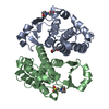
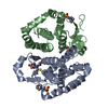
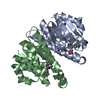
| Title | CRYSTAL STRUCTURE OF PI CLASS GLUTATHIONE TRANSFERASE | ||||||
 Components Components | GLUTATHIONE S-TRANSFERASE | ||||||
 Keywords Keywords | TRANSFERASE / glutathione transferase / helix capping mutant (S149A) | ||||||
| Function / homology |  Function and homology information Function and homology informationnitric oxide storage / S-nitrosoglutathione binding / TRAF2-GSTP1 complex / negative regulation of smooth muscle cell chemotaxis / negative regulation of leukocyte proliferation / dinitrosyl-iron complex binding / common myeloid progenitor cell proliferation / hepoxilin biosynthetic process / cellular response to cell-matrix adhesion / glutathione derivative biosynthetic process ...nitric oxide storage / S-nitrosoglutathione binding / TRAF2-GSTP1 complex / negative regulation of smooth muscle cell chemotaxis / negative regulation of leukocyte proliferation / dinitrosyl-iron complex binding / common myeloid progenitor cell proliferation / hepoxilin biosynthetic process / cellular response to cell-matrix adhesion / glutathione derivative biosynthetic process / response to L-ascorbic acid / linoleic acid metabolic process / Glutathione conjugation / nitric oxide binding / negative regulation of monocyte chemotactic protein-1 production / JUN kinase binding / glutathione peroxidase activity / Paracetamol ADME / oligodendrocyte development / negative regulation of stress-activated MAPK cascade / negative regulation of JNK cascade / cellular response to glucocorticoid stimulus / prostaglandin metabolic process / negative regulation of interleukin-1 beta production / regulation of stress-activated MAPK cascade / Detoxification of Reactive Oxygen Species / negative regulation of acute inflammatory response / glutathione transferase / negative regulation of ferroptosis / negative regulation of vascular associated smooth muscle cell proliferation / response to amino acid / glutathione transferase activity / animal organ regeneration / negative regulation of tumor necrosis factor production / protein serine/threonine kinase inhibitor activity / negative regulation of tumor necrosis factor-mediated signaling pathway / regulation of ERK1 and ERK2 cascade / toxic substance binding / negative regulation of fibroblast proliferation / positive regulation of superoxide anion generation / negative regulation of MAPK cascade / xenobiotic metabolic process / cellular response to epidermal growth factor stimulus / fatty acid binding / negative regulation of extrinsic apoptotic signaling pathway / response to reactive oxygen species / negative regulation of canonical NF-kappaB signal transduction / central nervous system development / glutathione metabolic process / negative regulation of ERK1 and ERK2 cascade / cellular response to insulin stimulus / response to estradiol / cellular response to lipopolysaccharide / secretory granule lumen / vesicle / ficolin-1-rich granule lumen / response to ethanol / Neutrophil degranulation / negative regulation of apoptotic process / negative regulation of transcription by RNA polymerase II / mitochondrion / : / extracellular exosome / extracellular region / nucleus / cytoplasm / cytosol Similarity search - Function | ||||||
| Biological species |  Homo sapiens (human) Homo sapiens (human) | ||||||
| Method |  X-RAY DIFFRACTION / Resolution: 2.1 Å X-RAY DIFFRACTION / Resolution: 2.1 Å | ||||||
 Authors Authors | Rossjohn, J. / McKinstry, W.J. / Oakley, A.J. / Parker, M.W. / Stenberg, G. / Mannervik, B. / Dragani, B. / Cocco, R. / Aceto, A. | ||||||
 Citation Citation | Journal: J.Mol.Biol. / Year: 2000 Title: Structures of thermolabile mutants of human glutathione transferase P1-1. Authors: Rossjohn, J. / McKinstry, W.J. / Oakley, A.J. / Parker, M.W. / Stenberg, G. / Mannervik, B. / Dragani, B. / Cocco, R. / Aceto, A. | ||||||
| History |
|
- Structure visualization
| Structure viewer | Molecule: MolmilJmol/JSmol |
|---|
- Downloads & links
-Download
| PDBx/mmCIF format | 1eog.cif.gz | 90.7 KB | Display | PDBx/mmCIF format |
|---|---|---|---|---|
| PDB format | pdb1eog.ent.gz | 70.2 KB | Display | PDB format |
| PDBx/mmJSON format | 1eog.json.gz | Tree view | PDBx/mmJSON format | |
| Others |  Other downloads Other downloads |
-Validation report
| Arichive directory | https://data.pdbj.org/pub/pdb/validation_reports/eo/1eogftp://data.pdbj.org/pub/pdb/validation_reports/eo/1eog | HTTPS FTP |
|---|
-Related structure data


-Links
 PDBj
PDBj


- Assembly
| Deposited unit | 
| ||||||||
|---|---|---|---|---|---|---|---|---|---|
| 1 |
| ||||||||
| Unit cell |
|
-Components
| #1: Protein | Mass: 23133.455 Da / Num. of mol.: 2 / Mutation: S149A Source method: isolated from a genetically manipulated source Source: (gene. exp.) Homo sapiens (human) / Plasmid: XL-1 BLUE / Organ (production host): LIVER / Production host:  #2: Water | ChemComp-HOH / |  Mass: 18.015 Da / Num. of mol.: 117 / Source method: isolated from a natural source / Formula: H2O Mass: 18.015 Da / Num. of mol.: 117 / Source method: isolated from a natural source / Formula: H2O |
|---|
-Experimental details
-Experiment
| Experiment | Method: X-RAY DIFFRACTION / Number of used crystals: 1 |
|---|
- Sample preparation
| Crystal | Density Matthews: 2.68 Å3/Da / Density % sol: 54.1 % | ||||||||||||||||||||||||||||||
|---|---|---|---|---|---|---|---|---|---|---|---|---|---|---|---|---|---|---|---|---|---|---|---|---|---|---|---|---|---|---|---|
| Crystal grow | Temperature: 293 K / Method: vapor diffusion, hanging drop / pH: 6.5 Details: 25% ammonium sulfate, pH 6.5, VAPOR DIFFUSION, HANGING DROP, temperature 20K | ||||||||||||||||||||||||||||||
| Crystal grow | *PLUS Temperature: 22 ℃ / pH: 7 | ||||||||||||||||||||||||||||||
| Components of the solutions | *PLUS
|
-Data collection
| Diffraction | Mean temperature: 100 K |
|---|---|
| Diffraction source | Source: ROTATING ANODE / Type: RIGAKU RU200 / Wavelength: 1.5418 |
| Detector | Type: MARRESEARCH / Detector: AREA DETECTOR / Date: Aug 1, 1999 |
| Radiation | Protocol: SINGLE WAVELENGTH / Monochromatic (M) / Laue (L): M / Scattering type: x-ray |
| Radiation wavelength | Wavelength: 1.5418 Å / Relative weight: 1 |
| Reflection | Resolution: 2.1→50 Å / Num. all: 26251 / Num. obs: 23250 |
| Reflection | *PLUS % possible obs: 91.6 % / Redundancy: 1.8 % / Num. measured all: 48527 / Rmerge(I) obs: 0.08 |
| Reflection shell | *PLUS Rmerge(I) obs: 0.361 / Mean I/σ(I) obs: 2 |
- Processing
| Software | Name: CNS / Classification: refinement | ||||||||||||||||||||||||
|---|---|---|---|---|---|---|---|---|---|---|---|---|---|---|---|---|---|---|---|---|---|---|---|---|---|
| Refinement | Resolution: 2.1→50 Å / σ(F): 0
| ||||||||||||||||||||||||
| Displacement parameters |
| ||||||||||||||||||||||||
| Refinement step | Cycle: LAST / Resolution: 2.1→50 Å
| ||||||||||||||||||||||||
| Refinement | *PLUS Rfactor Rfree: 0.238 / Rfactor Rwork: 0.204 | ||||||||||||||||||||||||
| Solvent computation | *PLUS | ||||||||||||||||||||||||
| Displacement parameters | *PLUS | ||||||||||||||||||||||||
| Refine LS restraints | *PLUS
|
