 Movie
Movie Controller

[English] 日本語
 Yorodumi
- PDB-1gqm: The structure of S100A12 in a hexameric form and its proposed rol... -
+ Open data

- Basic information
| Entry | Database: PDB / ID: 1gqm | ||||||
|---|---|---|---|---|---|---|---|
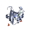
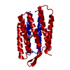
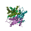
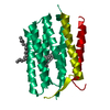
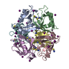
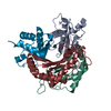
| Title | The structure of S100A12 in a hexameric form and its proposed role in receptor signalling | ||||||
 Components Components | CALGRANULIN C | ||||||
 Keywords Keywords | ANTIMICROBIAL PROTEIN / S100A12 / S100 PROTEIN FAMILY / EF-HAND / CALCIUM BINDING | ||||||
| Function / homology |  Function and homology information Function and homology informationmast cell activation / RAGE receptor binding / positive regulation of MAP kinase activity / TRAF6 mediated NF-kB activation / Advanced glycosylation endproduct receptor signaling / monocyte chemotaxis / defense response to fungus / endothelial cell migration / neutrophil chemotaxis / xenobiotic metabolic process ...mast cell activation / RAGE receptor binding / positive regulation of MAP kinase activity / TRAF6 mediated NF-kB activation / Advanced glycosylation endproduct receptor signaling / monocyte chemotaxis / defense response to fungus / endothelial cell migration / neutrophil chemotaxis / xenobiotic metabolic process / : / TAK1-dependent IKK and NF-kappa-B activation / calcium-dependent protein binding / positive regulation of inflammatory response / antimicrobial humoral immune response mediated by antimicrobial peptide / secretory granule lumen / killing of cells of another organism / cytoskeleton / positive regulation of canonical NF-kappaB signal transduction / defense response to bacterium / inflammatory response / copper ion binding / innate immune response / calcium ion binding / Neutrophil degranulation / extracellular region / zinc ion binding / identical protein binding / nucleus / plasma membrane / cytoplasm / cytosol Similarity search - Function | ||||||
| Biological species |  HOMO SAPIENS (human) HOMO SAPIENS (human) | ||||||
| Method |  X-RAY DIFFRACTION / SYNCHROTRON / MOLECULAR REPLACEMENT / Resolution: 2.7 Å X-RAY DIFFRACTION / SYNCHROTRON / MOLECULAR REPLACEMENT / Resolution: 2.7 Å | ||||||
 Authors Authors | Moroz, O.V. / Antson, A.A. / Dodson, E.G. / Burrel, H.J. / Grist, S.J. / Lloyd, R.M. / Maitland, N.J. / Dodson, G.G. / Wilson, K.S. / Lukanidin, E. / Bronstein, I.B. | ||||||
 Citation Citation | Journal: Acta Crystallogr.,Sect.D / Year: 2002 Title: The Structure of S100A12 in a Hexameric Form and its Proposed Role in Receptor Signalling Authors: Moroz, O.V. / Antson, A.A. / Dodson, E.J. / Burrell, H.J. / Grist, S.J. / Lloyd, R.M. / Maitland, N.J. / Dodson, G.G. / Wilson, K.S. / Lukanidin, E. / Bronstein, I.B. #1: Journal: Acta Crystallogr.,Sect.D / Year: 2001Title: The Three-Dimensional Structure of Human S100A12 Authors: Moroz, O.V. / Antson, A.A. / Murshudov, G.N. / Maitland, N.J. / Dodson, G.G. / Wilson, K.S. / Skibshoj, I. / Lukanidin, E.M. / Bronstein, I.B. #2: Journal: Acta Crystallogr.,Sect.D / Year: 2000 Title: Crystallization and Preliminary X-Ray Diffraction Analysis of Human Calcium-Binding Protein S100A12 Authors: Moroz, O.V. / Antson, A.A. / Dodson, G.G. / Wilson, K.S. / Skibshoj, I. / Lukanidin, E. / Bronstein, I. | ||||||
| History |
|
- Structure visualization
| Structure viewer | Molecule: MolmilJmol/JSmol |
|---|
- Downloads & links
-Download
| PDBx/mmCIF format | 1gqm.cif.gz | 228.6 KB | Display | PDBx/mmCIF format |
|---|---|---|---|---|
| PDB format | pdb1gqm.ent.gz | 179.1 KB | Display | PDB format |
| PDBx/mmJSON format | 1gqm.json.gz | Tree view | PDBx/mmJSON format | |
| Others |  Other downloads Other downloads |
-Validation report
| Arichive directory | https://data.pdbj.org/pub/pdb/validation_reports/gq/1gqmftp://data.pdbj.org/pub/pdb/validation_reports/gq/1gqm | HTTPS FTP |
|---|
-Related structure data
| Related structure data |  1e8aS S: Starting model for refinement |
|---|---|
| Similar structure data |


-Links
 PDBj
PDBj


- Assembly
| Deposited unit | 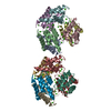
| |||||||||||||||||||||||||||||||||||||||||||||||||||||||||||||||||||||||||||||||||||||||||||||||||||||||||||||||||||||||||||||||||||||||||||||||||||||||||||||||||||||||||||||||||||||||||||||||||||||||||||||||||||||||||||||||||||||||||||||
|---|---|---|---|---|---|---|---|---|---|---|---|---|---|---|---|---|---|---|---|---|---|---|---|---|---|---|---|---|---|---|---|---|---|---|---|---|---|---|---|---|---|---|---|---|---|---|---|---|---|---|---|---|---|---|---|---|---|---|---|---|---|---|---|---|---|---|---|---|---|---|---|---|---|---|---|---|---|---|---|---|---|---|---|---|---|---|---|---|---|---|---|---|---|---|---|---|---|---|---|---|---|---|---|---|---|---|---|---|---|---|---|---|---|---|---|---|---|---|---|---|---|---|---|---|---|---|---|---|---|---|---|---|---|---|---|---|---|---|---|---|---|---|---|---|---|---|---|---|---|---|---|---|---|---|---|---|---|---|---|---|---|---|---|---|---|---|---|---|---|---|---|---|---|---|---|---|---|---|---|---|---|---|---|---|---|---|---|---|---|---|---|---|---|---|---|---|---|---|---|---|---|---|---|---|---|---|---|---|---|---|---|---|---|---|---|---|---|---|---|---|---|---|---|---|---|---|---|---|---|---|---|---|---|---|---|---|---|---|
| 1 | 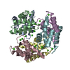
| |||||||||||||||||||||||||||||||||||||||||||||||||||||||||||||||||||||||||||||||||||||||||||||||||||||||||||||||||||||||||||||||||||||||||||||||||||||||||||||||||||||||||||||||||||||||||||||||||||||||||||||||||||||||||||||||||||||||||||||
| 2 | 
| |||||||||||||||||||||||||||||||||||||||||||||||||||||||||||||||||||||||||||||||||||||||||||||||||||||||||||||||||||||||||||||||||||||||||||||||||||||||||||||||||||||||||||||||||||||||||||||||||||||||||||||||||||||||||||||||||||||||||||||
| Unit cell |
| |||||||||||||||||||||||||||||||||||||||||||||||||||||||||||||||||||||||||||||||||||||||||||||||||||||||||||||||||||||||||||||||||||||||||||||||||||||||||||||||||||||||||||||||||||||||||||||||||||||||||||||||||||||||||||||||||||||||||||||
| Noncrystallographic symmetry (NCS) | NCS domain:
NCS domain segments:
NCS oper:
|
-Components
| #1: Protein | Mass: 10460.834 Da / Num. of mol.: 12 / Source method: isolated from a natural source / Source: (natural) HOMO SAPIENS (human) / Cell: GRANULOCYTE / Tissue: BLOOD / References: UniProt: P80511#2: Chemical | ChemComp-CA /   Mass: 40.078 Da / Num. of mol.: 36 / Source method: obtained synthetically / Formula: Ca Mass: 40.078 Da / Num. of mol.: 36 / Source method: obtained synthetically / Formula: Ca#3: Water | ChemComp-HOH / |  Mass: 18.015 Da / Num. of mol.: 26 / Source method: isolated from a natural source / Formula: H2O Mass: 18.015 Da / Num. of mol.: 26 / Source method: isolated from a natural source / Formula: H2O |
|---|
-Experimental details
-Experiment
| Experiment | Method: X-RAY DIFFRACTION / Number of used crystals: 1 |
|---|
- Sample preparation
| Crystal | Density Matthews: 2.4 Å3/Da / Density % sol: 48.9 % | |||||||||||||||||||||||||||||||||||
|---|---|---|---|---|---|---|---|---|---|---|---|---|---|---|---|---|---|---|---|---|---|---|---|---|---|---|---|---|---|---|---|---|---|---|---|---|
| Crystal grow | Method: vapor diffusion, hanging drop / pH: 6.5 Details: 20-25% PEG 5K MONOMETHYL ETHER, 0.2M CALCIUM CHLORIDE, 0.1M SODIUM CACODYLATE, HANGING DROP, pH 6.50 | |||||||||||||||||||||||||||||||||||
| Crystal grow | *PLUS Method: vapor diffusion, hanging drop | |||||||||||||||||||||||||||||||||||
| Components of the solutions | *PLUS
|
-Data collection
| Diffraction | Mean temperature: 120 K |
|---|---|
| Diffraction source | Source: SYNCHROTRON / Site: ESRF  / Beamline: ID14-4 / Wavelength: 0.93 / Beamline: ID14-4 / Wavelength: 0.93 |
| Detector | Date: Feb 15, 1999 |
| Radiation | Protocol: SINGLE WAVELENGTH / Monochromatic (M) / Laue (L): M / Scattering type: x-ray |
| Radiation wavelength | Wavelength: 0.93 Å / Relative weight: 1 |
| Reflection | Resolution: 2.7→20 Å / Num. obs: 32446 / % possible obs: 98.3 % / Redundancy: 2.6 % / Rmerge(I) obs: 0.053 / Net I/σ(I): 16.7 |
| Reflection shell | Resolution: 2.7→2.8 Å / Redundancy: 2.3 % / Rmerge(I) obs: 0.41 / Mean I/σ(I) obs: 2.3 / % possible all: 95.3 |
| Reflection shell | *PLUS Highest resolution: 2.7 Å / Lowest resolution: 2.8 Å / % possible obs: 95.3 % / Num. unique obs: 3107 / Rmerge(I) obs: 0.412 |
- Processing
| Software |
| ||||||||||||||||||||||||||||||||||||||||||||||||||||||||||||||||||||||||||||||||||||||||||||||||||||||||||||||||||||||||||||||||||||||||||||||||||||||||||||||||||||||||||||||||||||||
|---|---|---|---|---|---|---|---|---|---|---|---|---|---|---|---|---|---|---|---|---|---|---|---|---|---|---|---|---|---|---|---|---|---|---|---|---|---|---|---|---|---|---|---|---|---|---|---|---|---|---|---|---|---|---|---|---|---|---|---|---|---|---|---|---|---|---|---|---|---|---|---|---|---|---|---|---|---|---|---|---|---|---|---|---|---|---|---|---|---|---|---|---|---|---|---|---|---|---|---|---|---|---|---|---|---|---|---|---|---|---|---|---|---|---|---|---|---|---|---|---|---|---|---|---|---|---|---|---|---|---|---|---|---|---|---|---|---|---|---|---|---|---|---|---|---|---|---|---|---|---|---|---|---|---|---|---|---|---|---|---|---|---|---|---|---|---|---|---|---|---|---|---|---|---|---|---|---|---|---|---|---|---|---|
| Refinement | Method to determine structure: MOLECULAR REPLACEMENT Starting model: PDB ENTRY 1E8A Resolution: 2.7→19.84 Å / Cor.coef. Fo:Fc: 0.94 / Cor.coef. Fo:Fc free: 0.885 / SU B: 14.57 / SU ML: 0.302 / TLS residual ADP flag: LIKELY RESIDUAL / Cross valid method: THROUGHOUT / ESU R Free: 0.388 / Stereochemistry target values: MAXIMUM LIKELIHOOD
| ||||||||||||||||||||||||||||||||||||||||||||||||||||||||||||||||||||||||||||||||||||||||||||||||||||||||||||||||||||||||||||||||||||||||||||||||||||||||||||||||||||||||||||||||||||||
| Solvent computation | Ion probe radii: 0.8 Å / Shrinkage radii: 0.8 Å / VDW probe radii: 1.4 Å / Solvent model: BABINET MODEL WITH MASK | ||||||||||||||||||||||||||||||||||||||||||||||||||||||||||||||||||||||||||||||||||||||||||||||||||||||||||||||||||||||||||||||||||||||||||||||||||||||||||||||||||||||||||||||||||||||
| Refinement step | Cycle: LAST / Resolution: 2.7→19.84 Å
| ||||||||||||||||||||||||||||||||||||||||||||||||||||||||||||||||||||||||||||||||||||||||||||||||||||||||||||||||||||||||||||||||||||||||||||||||||||||||||||||||||||||||||||||||||||||
| Refine LS restraints |
|