 Movie
Movie Controller


+ Open data

- Basic information
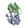
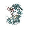
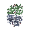
| Entry | Database: PDB / ID: 1gpe | |||||||||
|---|---|---|---|---|---|---|---|---|---|---|
| Title | GLUCOSE OXIDASE FROM PENICILLIUM AMAGASAKIENSE | |||||||||
 Components Components | PROTEIN (GLUCOSE OXIDASE) | |||||||||
 Keywords Keywords | OXIDOREDUCTASE(FLAVOPROTEIN) | |||||||||
| Function / homology |  Function and homology information Function and homology informationglucose oxidase / beta-D-glucose oxidase activity / flavin adenine dinucleotide binding / extracellular region / cytoplasm Similarity search - Function | |||||||||
| Biological species |  Penicillium amagasakiense (fungus) Penicillium amagasakiense (fungus) | |||||||||
| Method |  X-RAY DIFFRACTION / SYNCHROTRON / MOLECULAR REPLACEMENT / Resolution: 1.8 Å X-RAY DIFFRACTION / SYNCHROTRON / MOLECULAR REPLACEMENT / Resolution: 1.8 Å | |||||||||
 Authors Authors | Hendle, J. / Kalisz, H.M. / Hecht, H.J. | |||||||||
 Citation Citation | Journal: Acta Crystallogr.,Sect.D / Year: 1999 Title: 1.8 and 1.9 A resolution structures of the Penicillium amagasakiense and Aspergillus niger glucose oxidases as a basis for modelling substrate complexes. Authors: Wohlfahrt, G. / Witt, S. / Hendle, J. / Schomburg, D. / Kalisz, H.M. / Hecht, H.J. | |||||||||
| History |
|
- Structure visualization
| Structure viewer | Molecule: MolmilJmol/JSmol |
|---|
- Downloads & links
-Download
| PDBx/mmCIF format | 1gpe.cif.gz | 254.8 KB | Display | PDBx/mmCIF format |
|---|---|---|---|---|
| PDB format | pdb1gpe.ent.gz | 202.6 KB | Display | PDB format |
| PDBx/mmJSON format | 1gpe.json.gz | Tree view | PDBx/mmJSON format | |
| Others |  Other downloads Other downloads |
-Validation report
| Arichive directory | https://data.pdbj.org/pub/pdb/validation_reports/gp/1gpeftp://data.pdbj.org/pub/pdb/validation_reports/gp/1gpe | HTTPS FTP |
|---|
-Related structure data
| Related structure data | 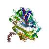 1cf3C 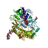 1galS S: Starting model for refinement C: citing same article ( |
|---|---|
| Similar structure data |


-Links
 PDBj
PDBj
- Assembly
| Deposited unit | 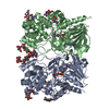
| ||||||||
|---|---|---|---|---|---|---|---|---|---|
| 1 |
| ||||||||
| Unit cell |
| ||||||||
| Noncrystallographic symmetry (NCS) | NCS oper: (Code: given Matrix: (-0.99995, 0.00686, -0.00701), Vector: |
-Components
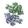
-Protein , 1 types, 2 molecules AB
| #1: Protein | Mass: 64016.750 Da / Num. of mol.: 2 / Source method: isolated from a natural source / Source: (natural) Penicillium amagasakiense (fungus) / References: UniProt: P81156, glucose oxidase |
|---|
-Sugars , 3 types, 8 molecules 
| #2: Polysaccharide | Source method: isolated from a genetically manipulated source #3: Polysaccharide | Source method: isolated from a genetically manipulated source #4: Sugar | ChemComp-NAG /  Type: D-saccharide, beta linking / Mass: 221.208 Da / Num. of mol.: 4 Type: D-saccharide, beta linking / Mass: 221.208 Da / Num. of mol.: 4Source method: isolated from a genetically manipulated source Formula: C8H15NO6 |
|---|
-Non-polymers , 2 types, 711 molecules 

| #5: Chemical |  Mass: 785.550 Da / Num. of mol.: 2 / Source method: obtained synthetically / Formula: C27H33N9O15P2 / Comment: FAD*YM Mass: 785.550 Da / Num. of mol.: 2 / Source method: obtained synthetically / Formula: C27H33N9O15P2 / Comment: FAD*YM#6: Water | ChemComp-HOH / | Mass: 18.015 Da / Num. of mol.: 709 / Source method: isolated from a natural source / Formula: H2O |
|---|
-Details
| Has protein modification | Y |
|---|
-Experimental details
-Experiment
| Experiment | Method: X-RAY DIFFRACTION / Number of used crystals: 1 |
|---|
- Sample preparation
| Crystal | Density Matthews: 2.21 Å3/Da / Density % sol: 44 % | ||||||||||||||||||||||||||||||||||||
|---|---|---|---|---|---|---|---|---|---|---|---|---|---|---|---|---|---|---|---|---|---|---|---|---|---|---|---|---|---|---|---|---|---|---|---|---|---|
| Crystal grow | pH: 7.4 Details: 1.3 M AMMONIUM SULPHATE, 0.1 M CITRATE-PO4 BUFFER PH 7.4 | ||||||||||||||||||||||||||||||||||||
| Crystal grow | *PLUS Method: vapor diffusion, hanging drop / Details: Kalisz, H.M., (1990) J.Mol.Biol., 213, 207. / PH range low: 5.6 / PH range high: 5.3 | ||||||||||||||||||||||||||||||||||||
| Components of the solutions | *PLUS
|
-Data collection
| Diffraction | Mean temperature: 290 K |
|---|---|
| Diffraction source | Source: SYNCHROTRON / Site: MPG/DESY, HAMBURG  / Beamline: BW6 / Wavelength: 1 / Beamline: BW6 / Wavelength: 1 |
| Detector | Type: MARRESEARCH / Detector: IMAGE PLATE / Details: MIRRORS |
| Radiation | Monochromator: SI(111) / Protocol: SINGLE WAVELENGTH / Monochromatic (M) / Laue (L): M / Scattering type: x-ray |
| Radiation wavelength | Wavelength: 1 Å / Relative weight: 1 |
| Reflection | Resolution: 1.8→20 Å / Num. obs: 101999 / % possible obs: 94.7 % / Observed criterion σ(I): 0 / Redundancy: 2.5 % / Biso Wilson estimate: 12.5 Å2 / Rmerge(I) obs: 0.068 / Net I/σ(I): 18.4 |
| Reflection shell | Resolution: 1.8→1.88 Å / Redundancy: 2 % / Rmerge(I) obs: 0.132 / Mean I/σ(I) obs: 8.5 / % possible all: 90 |
| Reflection shell | *PLUS Highest resolution: 1.8 Å / % possible obs: 90 % |
- Processing
| Software |
| ||||||||||||||||||||||||||||||||||||||||||||||||||||||||||||||||||||||||||||||||||||
|---|---|---|---|---|---|---|---|---|---|---|---|---|---|---|---|---|---|---|---|---|---|---|---|---|---|---|---|---|---|---|---|---|---|---|---|---|---|---|---|---|---|---|---|---|---|---|---|---|---|---|---|---|---|---|---|---|---|---|---|---|---|---|---|---|---|---|---|---|---|---|---|---|---|---|---|---|---|---|---|---|---|---|---|---|---|
| Refinement | Method to determine structure: MOLECULAR REPLACEMENT Starting model: PDB ENTRY 1GAL Resolution: 1.8→20 Å / Cross valid method: THROUGHOUT / σ(F): 0 / ESU R: 0.2
| ||||||||||||||||||||||||||||||||||||||||||||||||||||||||||||||||||||||||||||||||||||
| Displacement parameters | Biso mean: 14.9 Å2 | ||||||||||||||||||||||||||||||||||||||||||||||||||||||||||||||||||||||||||||||||||||
| Refinement step | Cycle: LAST / Resolution: 1.8→20 Å
| ||||||||||||||||||||||||||||||||||||||||||||||||||||||||||||||||||||||||||||||||||||
| Refine LS restraints |
| ||||||||||||||||||||||||||||||||||||||||||||||||||||||||||||||||||||||||||||||||||||
| Software | *PLUS Name: REFMAC / Classification: refinement | ||||||||||||||||||||||||||||||||||||||||||||||||||||||||||||||||||||||||||||||||||||
| Refinement | *PLUS Lowest resolution: 20 Å / % reflection Rfree: 5 % | ||||||||||||||||||||||||||||||||||||||||||||||||||||||||||||||||||||||||||||||||||||
| Solvent computation | *PLUS | ||||||||||||||||||||||||||||||||||||||||||||||||||||||||||||||||||||||||||||||||||||
| Displacement parameters | *PLUS | ||||||||||||||||||||||||||||||||||||||||||||||||||||||||||||||||||||||||||||||||||||
| Refine LS restraints | *PLUS
| ||||||||||||||||||||||||||||||||||||||||||||||||||||||||||||||||||||||||||||||||||||
| LS refinement shell | *PLUS Highest resolution: 1.8 Å / Lowest resolution: 1.88 Å / Rfactor Rfree: 0.237 / Rfactor obs: 0.179 |
