 Movie
Movie Controller


+ Open data

- Basic information
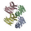
| Entry | Database: PDB / ID: 1gmw | ||||||
|---|---|---|---|---|---|---|---|
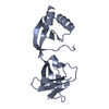
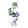
| Title | Structure of UreE | ||||||
 Components Components | (UREE) x 2 | ||||||
 Keywords Keywords | CHAPERONE / METALLOCHAPERONE | ||||||
| Function / homology |  Function and homology information Function and homology informationurea metabolic process / nickel cation binding / : / protein folding / protein-containing complex assembly / cytoplasm Similarity search - Function | ||||||
| Biological species |  KLEBSIELLA AEROGENES (bacteria) KLEBSIELLA AEROGENES (bacteria) | ||||||
| Method |  X-RAY DIFFRACTION / SYNCHROTRON / MOLECULAR REPLACEMENT / Resolution: 1.5 Å X-RAY DIFFRACTION / SYNCHROTRON / MOLECULAR REPLACEMENT / Resolution: 1.5 Å | ||||||
 Authors Authors | Song, H.K. / Mulrooney, S.B. / Huber, R. / Hausinger, R. | ||||||
 Citation Citation | Journal: J.Biol.Chem. / Year: 2001 Title: Crystal Structure of Klebsiella Aerogenes Uree, a Nickel-Binding Metallochaperone for Urease Activation Authors: Song, H.K. / Mulrooney, S.B. / Huber, R. / Hausinger, R. | ||||||
| History |
|
- Structure visualization
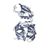
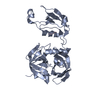
| Structure viewer | Molecule: MolmilJmol/JSmol |
|---|
- Downloads & links
-Download
| PDBx/mmCIF format | 1gmw.cif.gz | 120.9 KB | Display | PDBx/mmCIF format |
|---|---|---|---|---|
| PDB format | pdb1gmw.ent.gz | 95.7 KB | Display | PDB format |
| PDBx/mmJSON format | 1gmw.json.gz | Tree view | PDBx/mmJSON format | |
| Others |  Other downloads Other downloads |
-Validation report
| Arichive directory | https://data.pdbj.org/pub/pdb/validation_reports/gm/1gmwftp://data.pdbj.org/pub/pdb/validation_reports/gm/1gmw | HTTPS FTP |
|---|
-Related structure data
-Links
 PDBj
PDBj
- Assembly
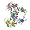
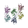
| Deposited unit | 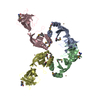
| ||||||||||||||||
|---|---|---|---|---|---|---|---|---|---|---|---|---|---|---|---|---|---|
| 1 |
| ||||||||||||||||
| Unit cell |
| ||||||||||||||||
| Noncrystallographic symmetry (NCS) | NCS oper:
|
-Components
| #1: Protein | Mass: 15924.580 Da / Num. of mol.: 3 / Fragment: RESIDUES 1-143 / Mutation: YES Source method: isolated from a genetically manipulated source Source: (gene. exp.) KLEBSIELLA AEROGENES (bacteria) / Production host: #2: Protein | | Mass: 15910.553 Da / Num. of mol.: 1 / Fragment: RESIDUES 1-143 / Mutation: YES Source method: isolated from a genetically manipulated source Source: (gene. exp.) KLEBSIELLA AEROGENES (bacteria) / Production host: #3: Chemical | ChemComp-CU /   Mass: 63.546 Da / Num. of mol.: 6 / Source method: obtained synthetically / Formula: Cu Mass: 63.546 Da / Num. of mol.: 6 / Source method: obtained synthetically / Formula: Cu#4: Water | ChemComp-HOH / |  Mass: 18.015 Da / Num. of mol.: 164 / Source method: isolated from a natural source / Formula: H2O Mass: 18.015 Da / Num. of mol.: 164 / Source method: isolated from a natural source / Formula: H2OCompound details | ENGINEERED | Has protein modification | Y | |
|---|
-Experimental details
-Experiment
| Experiment | Method: X-RAY DIFFRACTION / Number of used crystals: 1 |
|---|
- Sample preparation
| Crystal | Density Matthews: 2.52 Å3/Da / Density % sol: 51.14 % | ||||||||||||||||||||||||||||||||||||||||||||||||||||||
|---|---|---|---|---|---|---|---|---|---|---|---|---|---|---|---|---|---|---|---|---|---|---|---|---|---|---|---|---|---|---|---|---|---|---|---|---|---|---|---|---|---|---|---|---|---|---|---|---|---|---|---|---|---|---|---|
| Crystal grow | pH: 6.5 / Details: pH 6.50 | ||||||||||||||||||||||||||||||||||||||||||||||||||||||
| Crystal grow | *PLUS Temperature: 22 ℃ / pH: 7.5 / Method: vapor diffusion, hanging drop | ||||||||||||||||||||||||||||||||||||||||||||||||||||||
| Components of the solutions | *PLUS
|
-Data collection
| Diffraction | Mean temperature: 100 K |
|---|---|
| Diffraction source | Source: SYNCHROTRON / Site: MPG/DESY, HAMBURG  / Beamline: BW6 / Wavelength: 1.05 / Beamline: BW6 / Wavelength: 1.05 |
| Radiation | Protocol: SINGLE WAVELENGTH / Monochromatic (M) / Laue (L): M / Scattering type: x-ray |
| Radiation wavelength | Wavelength: 1.05 Å / Relative weight: 1 |
| Reflection | Resolution: 2.5→17 Å / Num. obs: 20709 / % possible obs: 97.5 % / Redundancy: 19 % / Rmerge(I) obs: 0.048 |
| Reflection | *PLUS Num. measured all: 198257 |
| Reflection shell | *PLUS Highest resolution: 2.5 Å / Lowest resolution: 2.54 Å / % possible obs: 99.2 % / Rmerge(I) obs: 0.088 |
- Processing
| Software | Name: CNS / Version: 1 / Classification: refinement | ||||||||||||||||||||||||||||||||||||||||||||||||||||||||||||
|---|---|---|---|---|---|---|---|---|---|---|---|---|---|---|---|---|---|---|---|---|---|---|---|---|---|---|---|---|---|---|---|---|---|---|---|---|---|---|---|---|---|---|---|---|---|---|---|---|---|---|---|---|---|---|---|---|---|---|---|---|---|
| Refinement | Method to determine structure: MOLECULAR REPLACEMENT / Resolution: 1.5→17 Å / Cross valid method: THROUGHOUT / σ(F): 0
| ||||||||||||||||||||||||||||||||||||||||||||||||||||||||||||
| Refinement step | Cycle: LAST / Resolution: 1.5→17 Å
| ||||||||||||||||||||||||||||||||||||||||||||||||||||||||||||
| Refine LS restraints |
| ||||||||||||||||||||||||||||||||||||||||||||||||||||||||||||
| Refinement | *PLUS Highest resolution: 2.5 Å / Lowest resolution: 17 Å / Num. reflection obs: 20352 / Rfactor Rfree: 0.295 | ||||||||||||||||||||||||||||||||||||||||||||||||||||||||||||
| Solvent computation | *PLUS | ||||||||||||||||||||||||||||||||||||||||||||||||||||||||||||
| Displacement parameters | *PLUS | ||||||||||||||||||||||||||||||||||||||||||||||||||||||||||||
| Refine LS restraints | *PLUS
|
