 Movie
Movie Controller


+ Open data

- Basic information
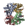
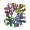
| Entry | Database: PDB / ID: 1gli | |||||||||
|---|---|---|---|---|---|---|---|---|---|---|
| Title | DEOXYHEMOGLOBIN T38W (ALPHA CHAINS), V1G (ALPHA AND BETA CHAINS) | |||||||||
 Components Components | (DEOXYHEMOGLOBIN) x 2 | |||||||||
 Keywords Keywords | OXYGEN TRANSPORT / MUTANT / ENGINEERED MUTANT / SITE DIRECTED MUTANT | |||||||||
| Function / homology |  Function and homology information Function and homology informationnitric oxide transport / hemoglobin alpha binding / cellular oxidant detoxification / hemoglobin binding / haptoglobin-hemoglobin complex / renal absorption / hemoglobin complex / oxygen transport / Scavenging of heme from plasma / endocytic vesicle lumen ...nitric oxide transport / hemoglobin alpha binding / cellular oxidant detoxification / hemoglobin binding / haptoglobin-hemoglobin complex / renal absorption / hemoglobin complex / oxygen transport / Scavenging of heme from plasma / endocytic vesicle lumen / blood vessel diameter maintenance / hydrogen peroxide catabolic process / oxygen carrier activity / carbon dioxide transport / response to hydrogen peroxide / Heme signaling / Erythrocytes take up oxygen and release carbon dioxide / Erythrocytes take up carbon dioxide and release oxygen / Late endosomal microautophagy / Cytoprotection by HMOX1 / oxygen binding / regulation of blood pressure / platelet aggregation / Chaperone Mediated Autophagy / positive regulation of nitric oxide biosynthetic process / tertiary granule lumen / Factors involved in megakaryocyte development and platelet production / blood microparticle / ficolin-1-rich granule lumen / iron ion binding / inflammatory response / heme binding / Neutrophil degranulation / extracellular space / extracellular exosome / extracellular region / metal ion binding / membrane / cytosol Similarity search - Function | |||||||||
| Biological species |  Homo sapiens (human) Homo sapiens (human) | |||||||||
| Method |  X-RAY DIFFRACTION / MUTANT-NATIVE DIFFERENCE MAP STARTING MODEL FOR MOLECULAR REPLACEMENT: NULL / Resolution: 2.5 Å X-RAY DIFFRACTION / MUTANT-NATIVE DIFFERENCE MAP STARTING MODEL FOR MOLECULAR REPLACEMENT: NULL / Resolution: 2.5 Å | |||||||||
 Authors Authors | Fermi, G. / Vallone, B. | |||||||||
 Citation Citation | Journal: J.Biol.Chem. / Year: 1996 Title: Probing the alpha 1 beta 2 interface of human hemoglobin by mutagenesis. Role of the FG-C contact regions. Authors: Vallone, B. / Bellelli, A. / Miele, A.E. / Brunori, M. / Fermi, G. | |||||||||
| History |
|
- Structure visualization
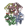
| Structure viewer | Molecule: MolmilJmol/JSmol |
|---|
- Downloads & links
-Download
| PDBx/mmCIF format | 1gli.cif.gz | 126.3 KB | Display | PDBx/mmCIF format |
|---|---|---|---|---|
| PDB format | pdb1gli.ent.gz | 100.3 KB | Display | PDB format |
| PDBx/mmJSON format | 1gli.json.gz | Tree view | PDBx/mmJSON format | |
| Others |  Other downloads Other downloads |
-Validation report
| Summary document | 1gli_validation.pdf.gz | 1.7 MB | Display | wwPDB validaton report |
|---|---|---|---|---|
| Full document | 1gli_full_validation.pdf.gz | 1.7 MB | Display | |
| Data in XML | 1gli_validation.xml.gz | 14.7 KB | Display | |
| Data in CIF | 1gli_validation.cif.gz | 19.9 KB | Display | |
| Arichive directory | https://data.pdbj.org/pub/pdb/validation_reports/gl/1gliftp://data.pdbj.org/pub/pdb/validation_reports/gl/1gli | HTTPS FTP |
-Related structure data
| Related structure data | |
|---|---|
| Similar structure data |


-Links
 PDBj
PDBj


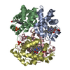
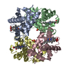
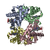
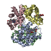
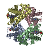
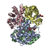
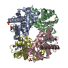
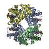
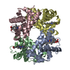
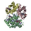
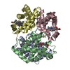
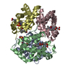
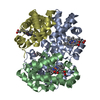
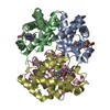
- Assembly
| Deposited unit | 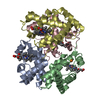
| ||||||||
|---|---|---|---|---|---|---|---|---|---|
| 1 |
| ||||||||
| Unit cell |
| ||||||||
| Details | THE CRYSTALLOGRAPHIC ASYMMETRIC UNIT CONTAINS TWO ALPHA AND TWO BETA CHAINS. ONLY ONE CHAIN OF EACH TYPE WAS DEPOSITED. THE COORDINATES FOR THE *C* AND *D* CHAINS WERE GENERATED FROM THE *A* AND *B* CHAINS, RESPECTIVELY, USING THE TRANSFORMATION (-X, Y, -Z). |
-Components
| #1: Protein | Mass: 15267.524 Da / Num. of mol.: 2 / Mutation: V1M, CHAIN A, C, T38W Source method: isolated from a genetically manipulated source Source: (gene. exp.) Homo sapiens (human) / Production host:  #2: Protein | Mass: 15922.265 Da / Num. of mol.: 2 / Mutation: V1M, CHAIN A, C, T38W Source method: isolated from a genetically manipulated source Source: (gene. exp.) Homo sapiens (human) / Production host: #3: Chemical | ChemComp-HEM /   Mass: 616.487 Da / Num. of mol.: 4 / Source method: obtained synthetically / Formula: C34H32FeN4O4 Mass: 616.487 Da / Num. of mol.: 4 / Source method: obtained synthetically / Formula: C34H32FeN4O4#4: Chemical | ChemComp-PO4 / |   Mass: 94.971 Da / Num. of mol.: 1 / Source method: obtained synthetically / Formula: PO4 Mass: 94.971 Da / Num. of mol.: 1 / Source method: obtained synthetically / Formula: PO4#5: Water | ChemComp-HOH / |  Mass: 18.015 Da / Num. of mol.: 110 / Source method: isolated from a natural source / Formula: H2O Mass: 18.015 Da / Num. of mol.: 110 / Source method: isolated from a natural source / Formula: H2OSequence details | THIS STRUCTURE IS AN ENGINEERED POINT MUTANT OF HUMAN HEMOGLOBIN, T38AW OR THR 38 ALPHA (1 AND 2) -- ...THIS STRUCTURE IS AN ENGINEERED | |
|---|
-Experimental details
-Experiment
| Experiment | Method: X-RAY DIFFRACTION / Number of used crystals: 1 |
|---|
- Sample preparation
| Crystal | Density Matthews: 2.25 Å3/Da / Density % sol: 50 % | ||||||||||||||||||||||||
|---|---|---|---|---|---|---|---|---|---|---|---|---|---|---|---|---|---|---|---|---|---|---|---|---|---|
| Crystal grow | *PLUS pH: 6.5 / Method: unknown / Details: Perutz, M.F., (1968) J. Cryst. Growth, 2, 54. | ||||||||||||||||||||||||
| Components of the solutions | *PLUS
|
-Data collection
| Diffraction | Mean temperature: 300 K |
|---|---|
| Diffraction source | Source: ROTATING ANODE / Wavelength: 1.5418 |
| Detector | Type: ENRAF-NONIUS FAST / Detector: DIFFRACTOMETER / Date: Aug 10, 1993 |
| Radiation | Monochromatic (M) / Laue (L): M / Scattering type: x-ray |
| Radiation wavelength | Wavelength: 1.5418 Å / Relative weight: 1 |
| Reflection | Resolution: 2.48→22 Å / Num. obs: 15818 / % possible obs: 76.5 % / Redundancy: 2 % / Rmerge(I) obs: 0.096 |
| Reflection | *PLUS Num. obs: 15560 / Num. measured all: 25572 |
- Processing
| Software |
| ||||||||||||
|---|---|---|---|---|---|---|---|---|---|---|---|---|---|
| Refinement | Method to determine structure: MUTANT-NATIVE DIFFERENCE MAP STARTING MODEL FOR MOLECULAR REPLACEMENT: NULL Highest resolution: 2.5 Å Details: THE COORDINATES GIVEN HERE ARE IN THE ORTHOGONAL ANGSTROM SYSTEM STANDARD FOR HEMOGLOBINS. THE Y AXIS IS THE (NON-CRYSTALLOGRAPHIC) MOLECULAR DIAD AND THE X AXIS IS THE PSEUDO-DIAD WHICH ...Details: THE COORDINATES GIVEN HERE ARE IN THE ORTHOGONAL ANGSTROM SYSTEM STANDARD FOR HEMOGLOBINS. THE Y AXIS IS THE (NON-CRYSTALLOGRAPHIC) MOLECULAR DIAD AND THE X AXIS IS THE PSEUDO-DIAD WHICH RELATES THE ALPHA-1 AND BETA-1 CHAINS. | ||||||||||||
| Refinement step | Cycle: LAST / Highest resolution: 2.5 Å
| ||||||||||||
| Refinement | *PLUS Num. reflection obs: 15442 / Rfactor obs: 0.135 | ||||||||||||
| Solvent computation | *PLUS | ||||||||||||
| Displacement parameters | *PLUS |
